

8
H. Chen et al.
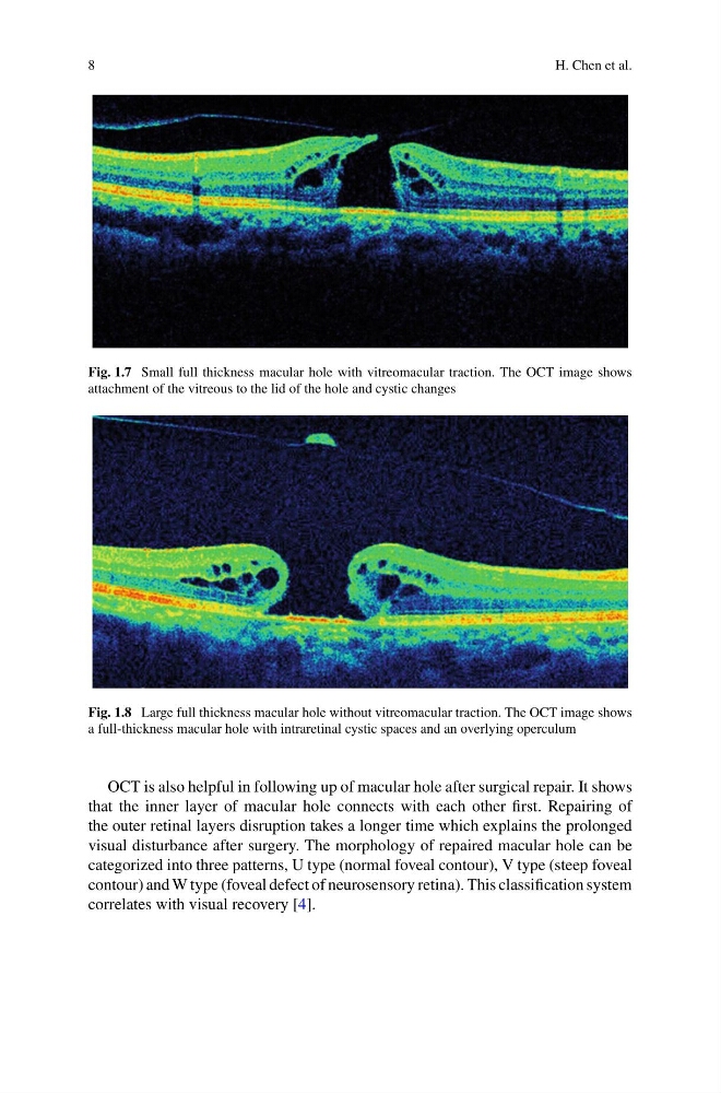
Fig. 1.7 Small full thickness macular hole with vitreomacular traction. The OCT image shows
attachment of the vitreous to the lid of the hole and cystic changes
Fig. 1.8 Large full thickness macular hole without vitreomacular traction. The OCT image shows
a full-thickness macular hole with intraretinal cystic spaces and an overlying operculum
OCT is also helpful in following up of macular hole after surgical repair. It shows
that the inner layer of macular hole connects with each other first. Repairing of
the outer retinal layers disruption takes a longer time which explains the prolonged
visual disturbance after surgery. The morphology of repaired macular hole can be
categorized into three patterns, U type (normal foveal contour), V type (steep foveal
contour) and W type (foveal defect of neurosensory retina). This classification system
correlates with visual recovery [4].
H. Chen et al.
Fig. 1.7 Small full thickness macular hole with vitreomacular traction. The OCT image shows
attachment of the vitreous to the lid of the hole and cystic changes
Fig. 1.8 Large full thickness macular hole without vitreomacular traction. The OCT image shows
a full-thickness macular hole with intraretinal cystic spaces and an overlying operculum
OCT is also helpful in following up of macular hole after surgical repair. It shows
that the inner layer of macular hole connects with each other first. Repairing of
the outer retinal layers disruption takes a longer time which explains the prolonged
visual disturbance after surgery. The morphology of repaired macular hole can be
categorized into three patterns, U type (normal foveal contour), V type (steep foveal
contour) and W type (foveal defect of neurosensory retina). This classification system
correlates with visual recovery [4].