

1 Clinical Applications of Retinal Optical Coherence Tomography
7
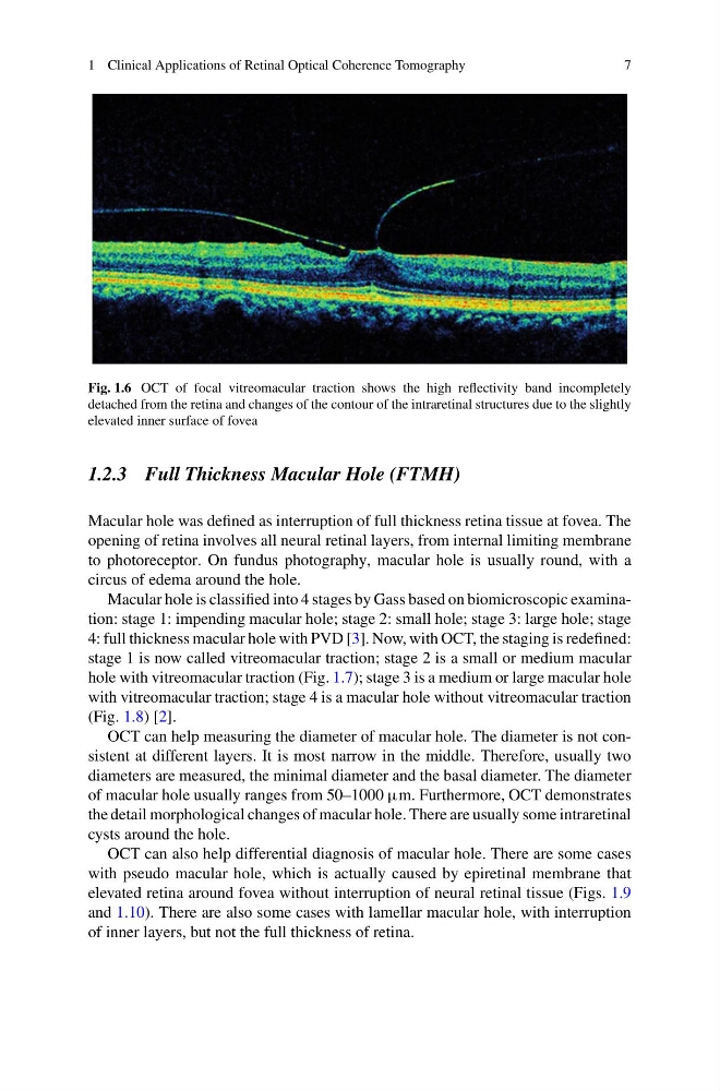
Fig. 1.6 OCT of focal vitreomacular traction shows the high reflectivity band incompletely
detached from the retina and changes of the contour of the intraretinal structures due to the slightly
elevated inner surface of fovea
1.2.3 Full Thickness Macular Hole (FTMH)
Macular hole was defined as interruption of full thickness retina tissue at fovea. The
opening of retina involves all neural retinal layers, from internal limiting membrane
to photoreceptor. On fundus photography, macular hole is usually round, with a
circus of edema around the hole.
Macular hole is classified into 4 stages by Gass based on biomicroscopic examination: stage 1: impending macular hole; stage 2: small hole; stage 3: large hole; stage
4: full thickness macular hole with PVD [3]. Now, with OCT, the staging is redefined:
stage 1 is now called vitreomacular traction; stage 2 is a small or medium macular
hole with vitreomacular traction (Fig. 1.7); stage 3 is a medium or large macular hole
with vitreomacular traction; stage 4 is a macular hole without vitreomacular traction
(Fig. 1.8) [2].
OCT can help measuring the diameter of macular hole. The diameter is not consistent at different layers. It is most narrow in the middle. Therefore, usually two
diameters are measured, the minimal diameter and the basal diameter. The diameter
of macular hole usually ranges from 50–1000 µm. Furthermore, OCT demonstrates
the detail morphological changes of macular hole. There are usually some intraretinal
cysts around the hole.
OCT can also help differential diagnosis of macular hole. There are some cases
with pseudo macular hole, which is actually caused by epiretinal membrane that
elevated retina around fovea without interruption of neural retinal tissue (Figs. 1.9
and 1.10). There are also some cases with lamellar macular hole, with interruption
of inner layers, but not the full thickness of retina.
7
Fig. 1.6 OCT of focal vitreomacular traction shows the high reflectivity band incompletely
detached from the retina and changes of the contour of the intraretinal structures due to the slightly
elevated inner surface of fovea
1.2.3 Full Thickness Macular Hole (FTMH)
Macular hole was defined as interruption of full thickness retina tissue at fovea. The
opening of retina involves all neural retinal layers, from internal limiting membrane
to photoreceptor. On fundus photography, macular hole is usually round, with a
circus of edema around the hole.
Macular hole is classified into 4 stages by Gass based on biomicroscopic examination: stage 1: impending macular hole; stage 2: small hole; stage 3: large hole; stage
4: full thickness macular hole with PVD [3]. Now, with OCT, the staging is redefined:
stage 1 is now called vitreomacular traction; stage 2 is a small or medium macular
hole with vitreomacular traction (Fig. 1.7); stage 3 is a medium or large macular hole
with vitreomacular traction; stage 4 is a macular hole without vitreomacular traction
(Fig. 1.8) [2].
OCT can help measuring the diameter of macular hole. The diameter is not consistent at different layers. It is most narrow in the middle. Therefore, usually two
diameters are measured, the minimal diameter and the basal diameter. The diameter
of macular hole usually ranges from 50–1000 µm. Furthermore, OCT demonstrates
the detail morphological changes of macular hole. There are usually some intraretinal
cysts around the hole.
OCT can also help differential diagnosis of macular hole. There are some cases
with pseudo macular hole, which is actually caused by epiretinal membrane that
elevated retina around fovea without interruption of neural retinal tissue (Figs. 1.9
and 1.10). There are also some cases with lamellar macular hole, with interruption
of inner layers, but not the full thickness of retina.