

6
H. Chen et al.
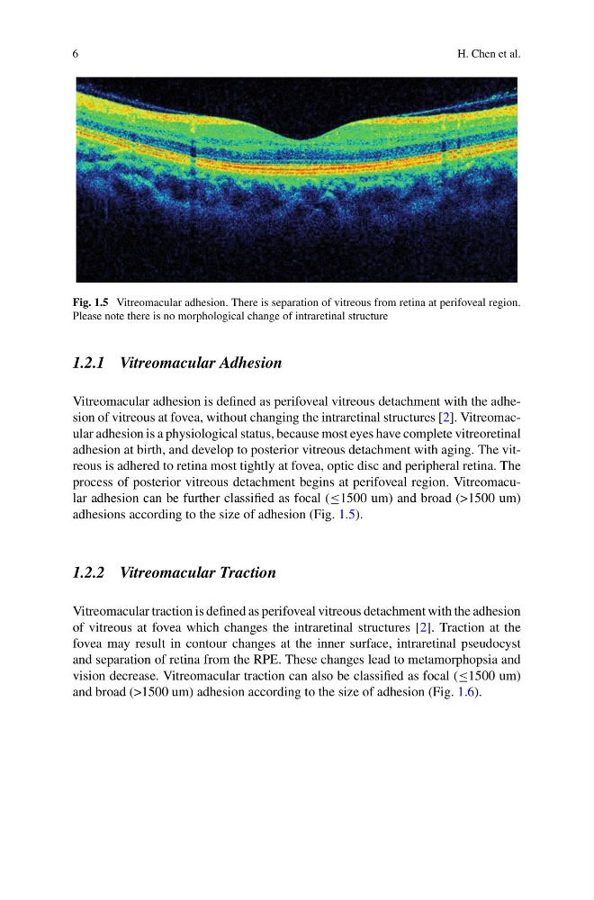
Fig. 1.5 Vitreomacular adhesion. There is separation of vitreous from retina at perifoveal region.
Please note there is no morphological change of intraretinal structure
1.2.1 Vitreomacular Adhesion
Vitreomacular adhesion is defined as perifoveal vitreous detachment with the adhesion of vitreous at fovea, without changing the intraretinal structures [2]. Vitreomacular adhesion is a physiological status, because most eyes have complete vitreoretinal
adhesion at birth, and develop to posterior vitreous detachment with aging. The vitreous is adhered to retina most tightly at fovea, optic disc and peripheral retina. The
process of posterior vitreous detachment begins at perifoveal region. Vitreomacular adhesion can be further classified as focal (≤1500 um) and broad (>1500 um)
adhesions according to the size of adhesion (Fig. 1.5).
1.2.2 Vitreomacular Traction
Vitreomacular traction is defined as perifoveal vitreous detachment with the adhesion
of vitreous at fovea which changes the intraretinal structures [2]. Traction at the
fovea may result in contour changes at the inner surface, intraretinal pseudocyst
and separation of retina from the RPE. These changes lead to metamorphopsia and
vision decrease. Vitreomacular traction can also be classified as focal (≤1500 um)
and broad (>1500 um) adhesion according to the size of adhesion (Fig. 1.6).
H. Chen et al.
Fig. 1.5 Vitreomacular adhesion. There is separation of vitreous from retina at perifoveal region.
Please note there is no morphological change of intraretinal structure
1.2.1 Vitreomacular Adhesion
Vitreomacular adhesion is defined as perifoveal vitreous detachment with the adhesion of vitreous at fovea, without changing the intraretinal structures [2]. Vitreomacular adhesion is a physiological status, because most eyes have complete vitreoretinal
adhesion at birth, and develop to posterior vitreous detachment with aging. The vitreous is adhered to retina most tightly at fovea, optic disc and peripheral retina. The
process of posterior vitreous detachment begins at perifoveal region. Vitreomacular adhesion can be further classified as focal (≤1500 um) and broad (>1500 um)
adhesions according to the size of adhesion (Fig. 1.5).
1.2.2 Vitreomacular Traction
Vitreomacular traction is defined as perifoveal vitreous detachment with the adhesion
of vitreous at fovea which changes the intraretinal structures [2]. Traction at the
fovea may result in contour changes at the inner surface, intraretinal pseudocyst
and separation of retina from the RPE. These changes lead to metamorphopsia and
vision decrease. Vitreomacular traction can also be classified as focal (≤1500 um)
and broad (>1500 um) adhesion according to the size of adhesion (Fig. 1.6).