

136
D. Cabrera DeBuc et al.
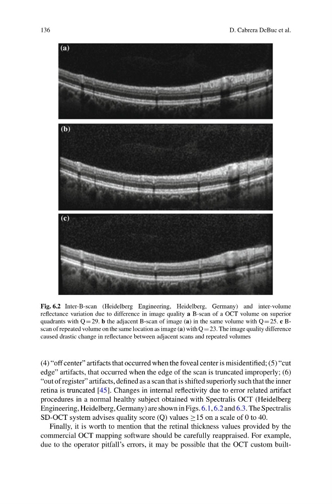
Fig. 6.2 Inter-B-scan (Heidelberg Engineering, Heidelberg, Germany) and inter-volume
reflectance variation due to difference in image quality a B-scan of a OCT volume on superior
quadrants with Q 29. b the adjacent B-scan of image (a) in the same volume with Q 25. c Bscan of repeated volume on the same location as image (a) with Q 23. The image quality difference
caused drastic change in reflectance between adjacent scans and repeated volumes
(4) “off center” artifacts that occurred when the foveal center is misidentified; (5) “cut
edge” artifacts, that occurred when the edge of the scan is truncated improperly; (6)
“out of register” artifacts, defined as a scan that is shifted superiorly such that the inner
retina is truncated [45]. Changes in internal reflectivity due to error related artifact
procedures in a normal healthy subject obtained with Spectralis OCT (Heidelberg
Engineering, Heidelberg, Germany) are shown in Figs. 6.1, 6.2 and 6.3. The Spectralis
SD-OCT system advises quality score (Q) values ≥15 on a scale of 0 to 40.
Finally, it is worth to mention that the retinal thickness values provided by the
commercial OCT mapping software should be carefully reappraised. For example,
due to the operator pitfall’s errors, it may be possible that the OCT custom built-
D. Cabrera DeBuc et al.
Fig. 6.2 Inter-B-scan (Heidelberg Engineering, Heidelberg, Germany) and inter-volume
reflectance variation due to difference in image quality a B-scan of a OCT volume on superior
quadrants with Q 29. b the adjacent B-scan of image (a) in the same volume with Q 25. c Bscan of repeated volume on the same location as image (a) with Q 23. The image quality difference
caused drastic change in reflectance between adjacent scans and repeated volumes
(4) “off center” artifacts that occurred when the foveal center is misidentified; (5) “cut
edge” artifacts, that occurred when the edge of the scan is truncated improperly; (6)
“out of register” artifacts, defined as a scan that is shifted superiorly such that the inner
retina is truncated [45]. Changes in internal reflectivity due to error related artifact
procedures in a normal healthy subject obtained with Spectralis OCT (Heidelberg
Engineering, Heidelberg, Germany) are shown in Figs. 6.1, 6.2 and 6.3. The Spectralis
SD-OCT system advises quality score (Q) values ≥15 on a scale of 0 to 40.
Finally, it is worth to mention that the retinal thickness values provided by the
commercial OCT mapping software should be carefully reappraised. For example,
due to the operator pitfall’s errors, it may be possible that the OCT custom built-