

6 Diagnostic Capability of Optical Coherence Tomography …
137
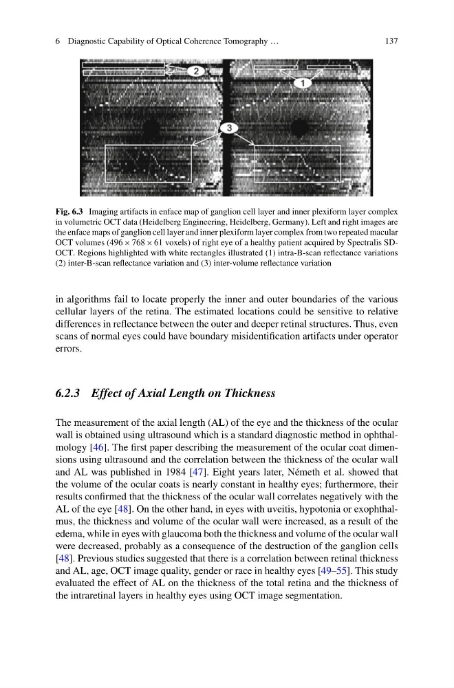
Fig. 6.3 Imaging artifacts in enface map of ganglion cell layer and inner plexiform layer complex
in volumetric OCT data (Heidelberg Engineering, Heidelberg, Germany). Left and right images are
the enface maps of ganglion cell layer and inner plexiform layer complex from two repeated macular
OCT volumes (496 × 768 × 61 voxels) of right eye of a healthy patient acquired by Spectralis SDOCT. Regions highlighted with white rectangles illustrated (1) intra-B-scan reflectance variations
(2) inter-B-scan reflectance variation and (3) inter-volume reflectance variation
in algorithms fail to locate properly the inner and outer boundaries of the various
cellular layers of the retina. The estimated locations could be sensitive to relative
differences in reflectance between the outer and deeper retinal structures. Thus, even
scans of normal eyes could have boundary misidentification artifacts under operator
errors.
6.2.3 Effect of Axial Length on Thickness
The measurement of the axial length (AL) of the eye and the thickness of the ocular
wall is obtained using ultrasound which is a standard diagnostic method in ophthalmology [46]. The first paper describing the measurement of the ocular coat dimensions using ultrasound and the correlation between the thickness of the ocular wall
and AL was published in 1984 [47]. Eight years later, Németh et al. showed that
the volume of the ocular coats is nearly constant in healthy eyes; furthermore, their
results confirmed that the thickness of the ocular wall correlates negatively with the
AL of the eye [48]. On the other hand, in eyes with uveitis, hypotonia or exophthalmus, the thickness and volume of the ocular wall were increased, as a result of the
edema, while in eyes with glaucoma both the thickness and volume of the ocular wall
were decreased, probably as a consequence of the destruction of the ganglion cells
[48]. Previous studies suggested that there is a correlation between retinal thickness
and AL, age, OCT image quality, gender or race in healthy eyes [49–55]. This study
evaluated the effect of AL on the thickness of the total retina and the thickness of
the intraretinal layers in healthy eyes using OCT image segmentation.
137
Fig. 6.3 Imaging artifacts in enface map of ganglion cell layer and inner plexiform layer complex
in volumetric OCT data (Heidelberg Engineering, Heidelberg, Germany). Left and right images are
the enface maps of ganglion cell layer and inner plexiform layer complex from two repeated macular
OCT volumes (496 × 768 × 61 voxels) of right eye of a healthy patient acquired by Spectralis SDOCT. Regions highlighted with white rectangles illustrated (1) intra-B-scan reflectance variations
(2) inter-B-scan reflectance variation and (3) inter-volume reflectance variation
in algorithms fail to locate properly the inner and outer boundaries of the various
cellular layers of the retina. The estimated locations could be sensitive to relative
differences in reflectance between the outer and deeper retinal structures. Thus, even
scans of normal eyes could have boundary misidentification artifacts under operator
errors.
6.2.3 Effect of Axial Length on Thickness
The measurement of the axial length (AL) of the eye and the thickness of the ocular
wall is obtained using ultrasound which is a standard diagnostic method in ophthalmology [46]. The first paper describing the measurement of the ocular coat dimensions using ultrasound and the correlation between the thickness of the ocular wall
and AL was published in 1984 [47]. Eight years later, Németh et al. showed that
the volume of the ocular coats is nearly constant in healthy eyes; furthermore, their
results confirmed that the thickness of the ocular wall correlates negatively with the
AL of the eye [48]. On the other hand, in eyes with uveitis, hypotonia or exophthalmus, the thickness and volume of the ocular wall were increased, as a result of the
edema, while in eyes with glaucoma both the thickness and volume of the ocular wall
were decreased, probably as a consequence of the destruction of the ganglion cells
[48]. Previous studies suggested that there is a correlation between retinal thickness
and AL, age, OCT image quality, gender or race in healthy eyes [49–55]. This study
evaluated the effect of AL on the thickness of the total retina and the thickness of
the intraretinal layers in healthy eyes using OCT image segmentation.