

120
F. Rathke et al.
(a) AMD
(b) DME
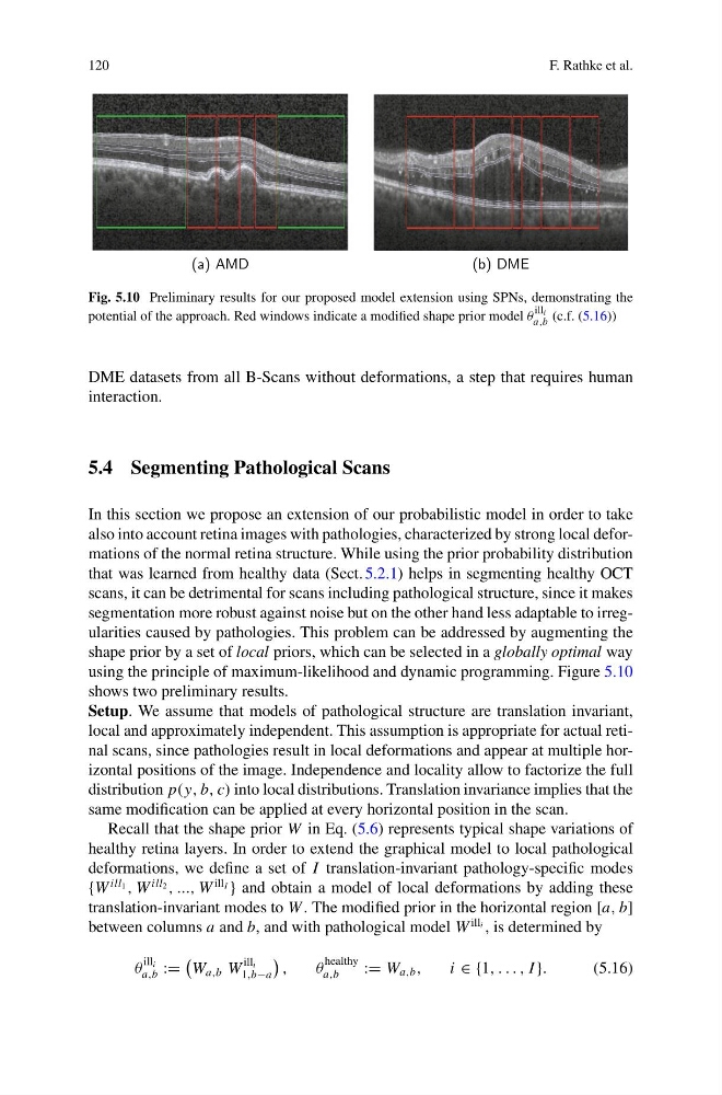
Fig. 5.10 Preliminary results for our proposed model extension using SPNs, demonstrating the
potential of the approach. Red windows indicate a modified shape prior model θ
ill i
a,b (c.f. (5.16))
DME datasets from all B-Scans without deformations, a step that requires human
interaction.
5.4 Segmenting Pathological Scans
In this section we propose an extension of our probabilistic model in order to take
also into account retina images with pathologies, characterized by strong local deformations of the normal retina structure. While using the prior probability distribution
that was learned from healthy data (Sect. 5.2.1) helps in segmenting healthy OCT
scans, it can be detrimental for scans including pathological structure, since it makes
segmentation more robust against noise but on the other hand less adaptable to irregularities caused by pathologies. This problem can be addressed by augmenting the
shape prior by a set of local priors, which can be selected in a globally optimal way
using the principle of maximum-likelihood and dynamic programming. Figure 5.10
shows two preliminary results.
Setup. We assume that models of pathological structure are translation invariant,
local and approximately independent. This assumption is appropriate for actual retinal scans, since pathologies result in local deformations and appear at multiple horizontal positions of the image. Independence and locality allow to factorize the full
distribution p(y, b, c) into local distributions. Translation invariance implies that the
same modification can be applied at every horizontal position in the scan.
Recall that the shape prior W in Eq. (5.6) represents typical shape variations of
healthy retina layers. In order to extend the graphical model to local pathological
deformations, we define a set of I translation-invariant pathology-specific modes
{W
ill 1 , W
ill 2 , ..., W
ill I } and obtain a model of local deformations by adding these
translation-invariant modes to W . The modified prior in the horizontal region [a, b]
between columns a and b, and with pathological model W
ill i , is determined by
θ
ill i
a,b :=
W a,b W
ill i
1,b−a
,
θ
healthy
a,b
:= W a,b ,
i ∈ {1, . . . , I }.
(5.16)
F. Rathke et al.
(a) AMD
(b) DME
Fig. 5.10 Preliminary results for our proposed model extension using SPNs, demonstrating the
potential of the approach. Red windows indicate a modified shape prior model θ
ill i
a,b (c.f. (5.16))
DME datasets from all B-Scans without deformations, a step that requires human
interaction.
5.4 Segmenting Pathological Scans
In this section we propose an extension of our probabilistic model in order to take
also into account retina images with pathologies, characterized by strong local deformations of the normal retina structure. While using the prior probability distribution
that was learned from healthy data (Sect. 5.2.1) helps in segmenting healthy OCT
scans, it can be detrimental for scans including pathological structure, since it makes
segmentation more robust against noise but on the other hand less adaptable to irregularities caused by pathologies. This problem can be addressed by augmenting the
shape prior by a set of local priors, which can be selected in a globally optimal way
using the principle of maximum-likelihood and dynamic programming. Figure 5.10
shows two preliminary results.
Setup. We assume that models of pathological structure are translation invariant,
local and approximately independent. This assumption is appropriate for actual retinal scans, since pathologies result in local deformations and appear at multiple horizontal positions of the image. Independence and locality allow to factorize the full
distribution p(y, b, c) into local distributions. Translation invariance implies that the
same modification can be applied at every horizontal position in the scan.
Recall that the shape prior W in Eq. (5.6) represents typical shape variations of
healthy retina layers. In order to extend the graphical model to local pathological
deformations, we define a set of I translation-invariant pathology-specific modes
{W
ill 1 , W
ill 2 , ..., W
ill I } and obtain a model of local deformations by adding these
translation-invariant modes to W . The modified prior in the horizontal region [a, b]
between columns a and b, and with pathological model W
ill i , is determined by
θ
ill i
a,b :=
W a,b W
ill i
1,b−a
,
θ
healthy
a,b
:= W a,b ,
i ∈ {1, . . . , I }.
(5.16)