

23
producing a high electron density. The schematic diagram of two glucopyranose
units of a cyclodextrin molecule showing details of the α-(1→4) glycosidic linkage
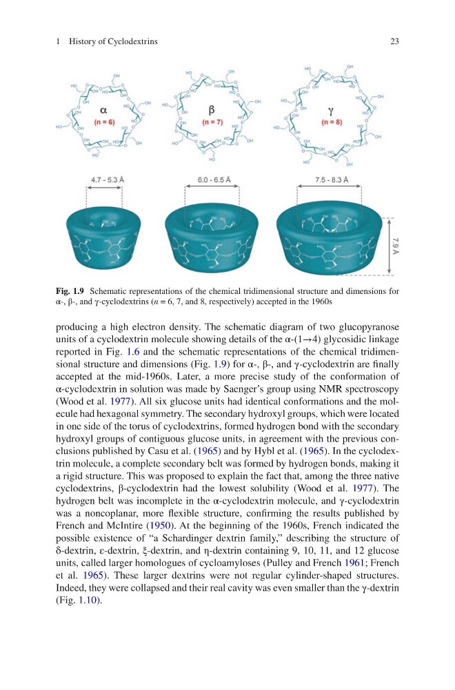
reported in Fig. 1.6 and the schematic representations of the chemical tridimensional structure and dimensions (Fig. 1.9) for α-, β-, and γ-cyclodextrin are finally
accepted at the mid-1960s. Later, a more precise study of the conformation of
α-cyclodextrin in solution was made by Saenger’s group using NMR spectroscopy
(Wood et al. 1977). All six glucose units had identical conformations and the molecule had hexagonal symmetry. The secondary hydroxyl groups, which were located
in one side of the torus of cyclodextrins, formed hydrogen bond with the secondary
hydroxyl groups of contiguous glucose units, in agreement with the previous conclusions published by Casu et al. (1965) and by Hybl et al. (1965). In the cyclodextrin molecule, a complete secondary belt was formed by hydrogen bonds, making it
a rigid structure. This was proposed to explain the fact that, among the three native
cyclodextrins, β-cyclodextrin had the lowest solubility (Wood et al. 1977). The
hydrogen belt was incomplete in the α-cyclodextrin molecule, and γ-cyclodextrin
was a noncoplanar, more flexible structure, confirming the results published by
French and McIntire (1950). At the beginning of the 1960s, French indicated the
possible existence of “a Schardinger dextrin family,” describing the structure of
δ-dextrin, ε-dextrin, ξ-dextrin, and η-dextrin containing 9, 10, 11, and 12 glucose
units, called larger homologues of cycloamyloses (Pulley and French 1961; French
et al. 1965). These larger dextrins were not regular cylinder-shaped structures.
Indeed, they were collapsed and their real cavity was even smaller than the γ-dextrin
(Fig. 1.10).
Fig. 1.9 Schematic representations of the chemical tridimensional structure and dimensions for
α-, β-, and γ-cyclodextrins (n = 6, 7, and 8, respectively) accepted in the 1960s
1 History of Cyclodextrins
producing a high electron density. The schematic diagram of two glucopyranose
units of a cyclodextrin molecule showing details of the α-(1→4) glycosidic linkage
reported in Fig. 1.6 and the schematic representations of the chemical tridimensional structure and dimensions (Fig. 1.9) for α-, β-, and γ-cyclodextrin are finally
accepted at the mid-1960s. Later, a more precise study of the conformation of
α-cyclodextrin in solution was made by Saenger’s group using NMR spectroscopy
(Wood et al. 1977). All six glucose units had identical conformations and the molecule had hexagonal symmetry. The secondary hydroxyl groups, which were located
in one side of the torus of cyclodextrins, formed hydrogen bond with the secondary
hydroxyl groups of contiguous glucose units, in agreement with the previous conclusions published by Casu et al. (1965) and by Hybl et al. (1965). In the cyclodextrin molecule, a complete secondary belt was formed by hydrogen bonds, making it
a rigid structure. This was proposed to explain the fact that, among the three native
cyclodextrins, β-cyclodextrin had the lowest solubility (Wood et al. 1977). The
hydrogen belt was incomplete in the α-cyclodextrin molecule, and γ-cyclodextrin
was a noncoplanar, more flexible structure, confirming the results published by
French and McIntire (1950). At the beginning of the 1960s, French indicated the
possible existence of “a Schardinger dextrin family,” describing the structure of
δ-dextrin, ε-dextrin, ξ-dextrin, and η-dextrin containing 9, 10, 11, and 12 glucose
units, called larger homologues of cycloamyloses (Pulley and French 1961; French
et al. 1965). These larger dextrins were not regular cylinder-shaped structures.
Indeed, they were collapsed and their real cavity was even smaller than the γ-dextrin
(Fig. 1.10).
Fig. 1.9 Schematic representations of the chemical tridimensional structure and dimensions for
α-, β-, and γ-cyclodextrins (n = 6, 7, and 8, respectively) accepted in the 1960s
1 History of Cyclodextrins