

22
features, using NMR spectra in dimethylsulfoxide solution and using X-ray crystallography of the α-cyclodextrin-potassium acetate complex, respectively. Their
results clearly demonstrated that (i) all the glucose residues of cyclodextrins were in
the
4
C1 chair conformation; (ii) the cavity was lined by the hydrogen atoms and the
glycosidic oxygen bridges, respectively; and (iii) the nonbonding electron pairs of
the glycosidic oxygen bridges were directed toward the inside of the cavity,
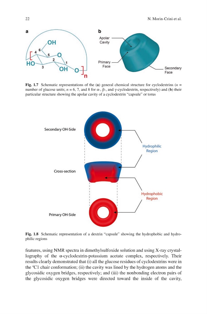
Fig. 1.8 Schematic representation of a dextrin “capsule” showing the hydrophobic and hydrophilic regions
Fig. 1.7 Schematic representations of the (a) general chemical structure for cyclodextrins (n =
number of glucose units; n = 6, 7, and 8 for α-, β-, and γ-cyclodextrin, respectively) and (b) their
particular structure showing the apolar cavity of a cyclodextrin “capsule” or torus
N. Morin-Crini et al.
features, using NMR spectra in dimethylsulfoxide solution and using X-ray crystallography of the α-cyclodextrin-potassium acetate complex, respectively. Their
results clearly demonstrated that (i) all the glucose residues of cyclodextrins were in
the
4
C1 chair conformation; (ii) the cavity was lined by the hydrogen atoms and the
glycosidic oxygen bridges, respectively; and (iii) the nonbonding electron pairs of
the glycosidic oxygen bridges were directed toward the inside of the cavity,
Fig. 1.8 Schematic representation of a dextrin “capsule” showing the hydrophobic and hydrophilic regions
Fig. 1.7 Schematic representations of the (a) general chemical structure for cyclodextrins (n =
number of glucose units; n = 6, 7, and 8 for α-, β-, and γ-cyclodextrin, respectively) and (b) their
particular structure showing the apolar cavity of a cyclodextrin “capsule” or torus
N. Morin-Crini et al.