

120
As a consequence of the C-1 conformation of the glucopyranose units, all secondary hydroxyl groups were situated on one of the two edges of the ring, whereas
all the primary ones were placed on the other edge. The cavity, composed of several
glucose units, was lined by the hydrogen atoms and the glycosidic oxygen bridges,
respectively. The nonbonding electron pairs of the glycosidic oxygen bridges were
directed toward the inside of the cavity producing there a high electron density and
lending it some Lewis-base character. A schematic representation of a “cyclodextrin
capsule” is reported in Fig. 2.14.
Like Professor French, Professor Szejtli pointed out the fact that a cyclodextrin
molecule should be regarded rather as a truncated cone than a cylinder. The core of
this structure can trap or encapsulate other substances (Szejtli 1995). Figure 2.15
illustrates the hydrophilic and hydrophobic regions of cyclodextrins.
Using the results of Professor Casu on the chemical structure of cyclodextrins
obtained from infrared and NMR experiments, Professor Szejtli also indicated that
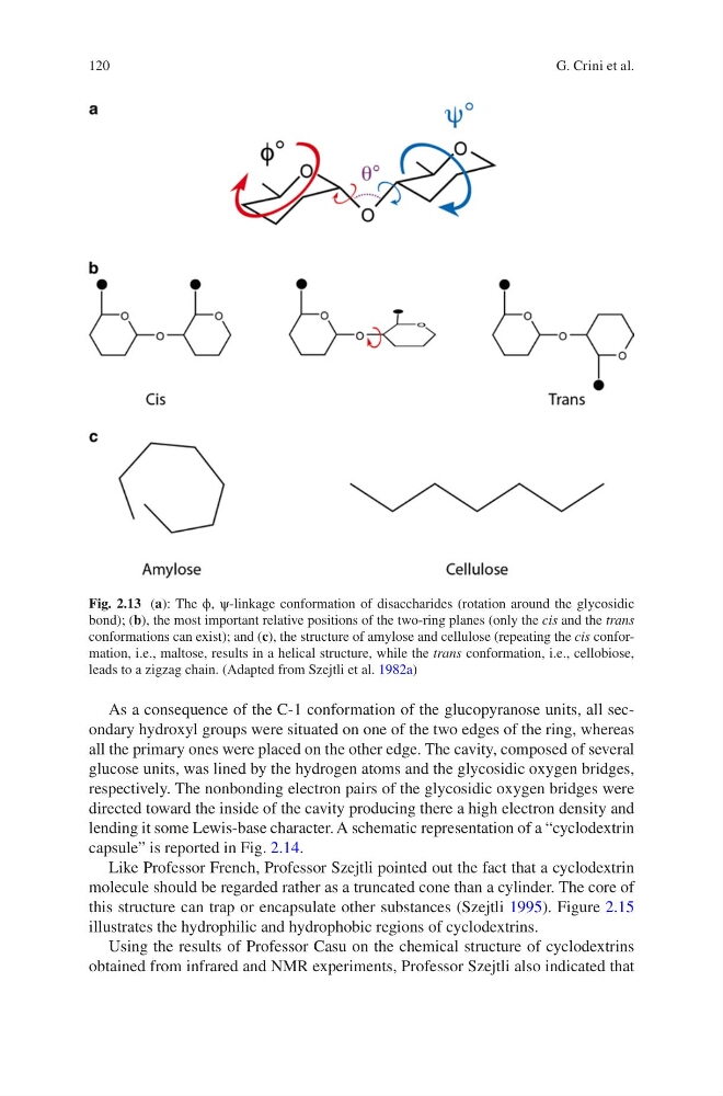
Fig. 2.13 (a): The ϕ, ψ-linkage conformation of disaccharides (rotation around the glycosidic
bond); (b), the most important relative positions of the two-ring planes (only the cis and the trans
conformations can exist); and (c), the structure of amylose and cellulose (repeating the cis conformation, i.e., maltose, results in a helical structure, while the trans conformation, i.e., cellobiose,
leads to a zigzag chain. (Adapted from Szejtli et al. 1982a)
G. Crini et al.
As a consequence of the C-1 conformation of the glucopyranose units, all secondary hydroxyl groups were situated on one of the two edges of the ring, whereas
all the primary ones were placed on the other edge. The cavity, composed of several
glucose units, was lined by the hydrogen atoms and the glycosidic oxygen bridges,
respectively. The nonbonding electron pairs of the glycosidic oxygen bridges were
directed toward the inside of the cavity producing there a high electron density and
lending it some Lewis-base character. A schematic representation of a “cyclodextrin
capsule” is reported in Fig. 2.14.
Like Professor French, Professor Szejtli pointed out the fact that a cyclodextrin
molecule should be regarded rather as a truncated cone than a cylinder. The core of
this structure can trap or encapsulate other substances (Szejtli 1995). Figure 2.15
illustrates the hydrophilic and hydrophobic regions of cyclodextrins.
Using the results of Professor Casu on the chemical structure of cyclodextrins
obtained from infrared and NMR experiments, Professor Szejtli also indicated that
Fig. 2.13 (a): The ϕ, ψ-linkage conformation of disaccharides (rotation around the glycosidic
bond); (b), the most important relative positions of the two-ring planes (only the cis and the trans
conformations can exist); and (c), the structure of amylose and cellulose (repeating the cis conformation, i.e., maltose, results in a helical structure, while the trans conformation, i.e., cellobiose,
leads to a zigzag chain. (Adapted from Szejtli et al. 1982a)
G. Crini et al.