

1 3
Top Curr Chem (Z) (2018) 376:24
and brightness of the CT states are affected by inter-chromophore distances, their
π-orbital overlaps and environmental effects explicitly accounted for in our SOS//
QM/MM approach.
Considering the benchmark study of the protein aromatic chromophores
described in the previous section, which provides converged results that are consistent with available experimental data, we can conclude that our predictions for
two-dimensional electronic spectra strongly support the use of the 2DUV technique
as an analytical tool for monitoring GS conformational dynamics of protein systems
containing aromatic UV-chromophores.
We expect that the adoption of 2DUV will be equally well suited for tracking
of GS conformational changes in nucleic acids. Dinucleoside monophosphates,
i.e. dimers of DNA/RNA nucleobases, represent excellent model systems for
exploring this hypothesis from our theoretical perspective. In fact, they are realistic biomolecules that are treatable by our QM approaches [57, 60] and assume
various stacking conformations in water solution [85], currently representing
model systems for studying the complex excited-state dynamics of nucleic acids
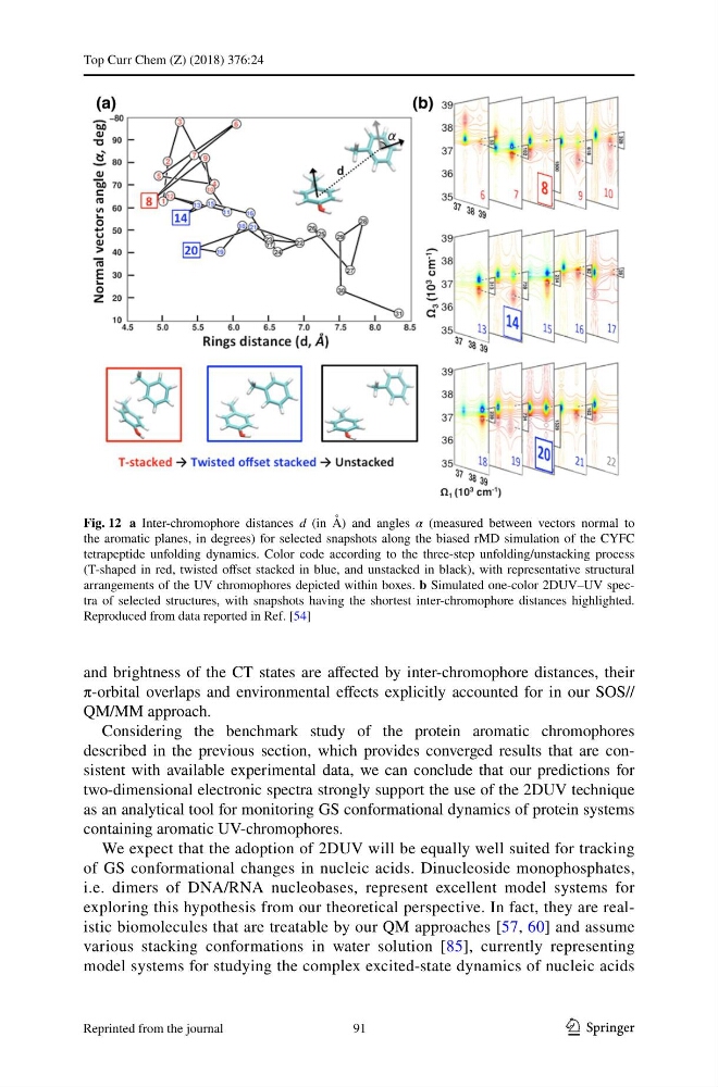
Fig. 12 a Inter-chromophore distances d (in Å) and angles α (measured between vectors normal to
the aromatic planes, in degrees) for selected snapshots along the biased rMD simulation of the CYFC
tetrapeptide unfolding dynamics. Color code according to the three-step unfolding/unstacking process
(T-shaped in red, twisted offset stacked in blue, and unstacked in black), with representative structural
arrangements of the UV chromophores depicted within boxes. b Simulated one-color 2DUV–UV spectra of selected structures, with snapshots having the shortest inter-chromophore distances highlighted.
Reproduced from data reported in Ref. [54]
91
Reprinted from the journal
Top Curr Chem (Z) (2018) 376:24
and brightness of the CT states are affected by inter-chromophore distances, their
π-orbital overlaps and environmental effects explicitly accounted for in our SOS//
QM/MM approach.
Considering the benchmark study of the protein aromatic chromophores
described in the previous section, which provides converged results that are consistent with available experimental data, we can conclude that our predictions for
two-dimensional electronic spectra strongly support the use of the 2DUV technique
as an analytical tool for monitoring GS conformational dynamics of protein systems
containing aromatic UV-chromophores.
We expect that the adoption of 2DUV will be equally well suited for tracking
of GS conformational changes in nucleic acids. Dinucleoside monophosphates,
i.e. dimers of DNA/RNA nucleobases, represent excellent model systems for
exploring this hypothesis from our theoretical perspective. In fact, they are realistic biomolecules that are treatable by our QM approaches [57, 60] and assume
various stacking conformations in water solution [85], currently representing
model systems for studying the complex excited-state dynamics of nucleic acids
Fig. 12 a Inter-chromophore distances d (in Å) and angles α (measured between vectors normal to
the aromatic planes, in degrees) for selected snapshots along the biased rMD simulation of the CYFC
tetrapeptide unfolding dynamics. Color code according to the three-step unfolding/unstacking process
(T-shaped in red, twisted offset stacked in blue, and unstacked in black), with representative structural
arrangements of the UV chromophores depicted within boxes. b Simulated one-color 2DUV–UV spectra of selected structures, with snapshots having the shortest inter-chromophore distances highlighted.
Reproduced from data reported in Ref. [54]
91
Reprinted from the journal