

1 H NMR spectrum of the complex provides valuable information on the structure. This is illustrated by taking complex 28 as an example. Figure 3 depicts the
1 H NMR spectra with the assignment. The i proton appears broad, and it shows a
downfield shift (Δδ = 0.57 ppm, for 28) relative to the free ligand. This indicates
preagostic interaction of proton i with Ru. The a
0 proton undergoes a large upfield
shift relative to the free ligand (Δδ = 1.91 ppm). The a
0 proton is located over the
phenyl ring of the second ligand. A large upfield shift is attributed to the diamagnetic shielding of the aromatic ring from the second ligand. Similar observations were made for M–M bonded dirhodium(II,II) and dimolybdenum(II,II)
complexes [101].
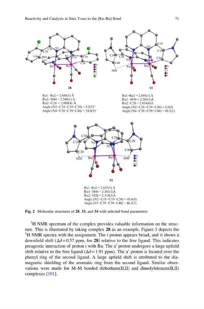
28
Ru1−Ru2 = 2.688(1) Å
Ru1−H44 = 2.540(1) Å
Ru2−C24 = 2.068(4) Å
Angle (N2−C18−C19−C24) = 5.2(5)°
Angle (N4−C38−C39−C44) = 54.0(5)°
33
Ru1−Ru2 = 2.695(1) Å
Ru1−H40 = 2.503(1)Å
Ru2−C20 = 2.054(6)Å
Angle (N2−C18−C19−C20) = 2.8(9)
Angle (N4−C38−C39−C40) = 49.2(1)
34
Ru1−Ru2 = 2.655(1) Å
Ru1−H40 = 2.301(1)Å
Ru2−H20 = 2.314(1)Å
Angle (N2−C18−C19−C20) = 45.6(9)
Angle (N5−C38−C39−C40) = 46.2(7)
Fig. 2 Molecular structures of 28, 33, and 34 with selected bond parameters
Reactivity and Catalysis at Sites Trans to the [Ru–Ru] Bond
71
1 H NMR spectra with the assignment. The i proton appears broad, and it shows a
downfield shift (Δδ = 0.57 ppm, for 28) relative to the free ligand. This indicates
preagostic interaction of proton i with Ru. The a
0 proton undergoes a large upfield
shift relative to the free ligand (Δδ = 1.91 ppm). The a
0 proton is located over the
phenyl ring of the second ligand. A large upfield shift is attributed to the diamagnetic shielding of the aromatic ring from the second ligand. Similar observations were made for M–M bonded dirhodium(II,II) and dimolybdenum(II,II)
complexes [101].
28
Ru1−Ru2 = 2.688(1) Å
Ru1−H44 = 2.540(1) Å
Ru2−C24 = 2.068(4) Å
Angle (N2−C18−C19−C24) = 5.2(5)°
Angle (N4−C38−C39−C44) = 54.0(5)°
33
Ru1−Ru2 = 2.695(1) Å
Ru1−H40 = 2.503(1)Å
Ru2−C20 = 2.054(6)Å
Angle (N2−C18−C19−C20) = 2.8(9)
Angle (N4−C38−C39−C40) = 49.2(1)
34
Ru1−Ru2 = 2.655(1) Å
Ru1−H40 = 2.301(1)Å
Ru2−H20 = 2.314(1)Å
Angle (N2−C18−C19−C20) = 45.6(9)
Angle (N5−C38−C39−C40) = 46.2(7)
Fig. 2 Molecular structures of 28, 33, and 34 with selected bond parameters
Reactivity and Catalysis at Sites Trans to the [Ru–Ru] Bond
71