

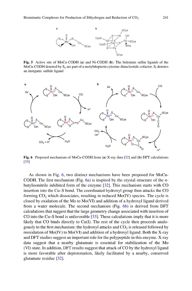
As shown in Fig. 6, two distinct mechanisms have been proposed for MoCuCODH. The first mechanism (Fig. 6a) is inspired by the crystal structure of the nbutylisonitrile inhibited form of the enzyme [32]. This mechanism starts with CO
insertion into the Cu–S bond. The coordinated hydroxyl group then attacks the CO
forming CO 2 which dissociates, resulting in reduced Mo(IV) species. The cycle is
closed by oxidation of the Mo to Mo(VI) and addition of a hydroxyl ligand derived
from a water molecule. The second mechanism (Fig. 6b) is derived from DFT
calculations that suggest that the large geometry change associated with insertion of
CO into the Cu–S bond is unfavorable [33]. These calculations imply that it is more
likely that CO binds directly to Cu(I). The rest of the cycle then proceeds analogously to the first mechanism: the hydroxyl attacks and CO 2 is released followed by
reoxidation of Mo(IV) to Mo(VI) and addition of a hydroxyl ligand. Both the X-ray
and DFT studies suggest an important role for the polypeptide in this enzyme. X-ray
data suggest that a nearby glutamate is essential for stabilization of the Mo
(VI) state. In addition, DFT results suggest that attack of CO by the hydroxyl ligand
is more favorable after deprotonation, likely facilitated by a nearby, conserved
glutamate residue [32].
Fig. 5 Active site of MoCu-CODH (a) and Ni-CODH (b). The bidentate sulfur ligands of the
MoCu-CODH denoted by S o are part of a molybdopterin cytosine dinucleotide cofactor. S i denotes
an inorganic sulfide ligand
Fig. 6 Proposed mechanism of MoCu-CODH from (a) X-ray data [32] and (b) DFT calculations
[33]
Biomimetic Complexes for Production of Dihydrogen and Reduction of CO 2
241
insertion into the Cu–S bond. The coordinated hydroxyl group then attacks the CO
forming CO 2 which dissociates, resulting in reduced Mo(IV) species. The cycle is
closed by oxidation of the Mo to Mo(VI) and addition of a hydroxyl ligand derived
from a water molecule. The second mechanism (Fig. 6b) is derived from DFT
calculations that suggest that the large geometry change associated with insertion of
CO into the Cu–S bond is unfavorable [33]. These calculations imply that it is more
likely that CO binds directly to Cu(I). The rest of the cycle then proceeds analogously to the first mechanism: the hydroxyl attacks and CO 2 is released followed by
reoxidation of Mo(IV) to Mo(VI) and addition of a hydroxyl ligand. Both the X-ray
and DFT studies suggest an important role for the polypeptide in this enzyme. X-ray
data suggest that a nearby glutamate is essential for stabilization of the Mo
(VI) state. In addition, DFT results suggest that attack of CO by the hydroxyl ligand
is more favorable after deprotonation, likely facilitated by a nearby, conserved
glutamate residue [32].
Fig. 5 Active site of MoCu-CODH (a) and Ni-CODH (b). The bidentate sulfur ligands of the
MoCu-CODH denoted by S o are part of a molybdopterin cytosine dinucleotide cofactor. S i denotes
an inorganic sulfide ligand
Fig. 6 Proposed mechanism of MoCu-CODH from (a) X-ray data [32] and (b) DFT calculations
[33]
Biomimetic Complexes for Production of Dihydrogen and Reduction of CO 2
241