

pinpoint spatial accuracy also makes SXRS a convenient tool in studying different
ET pathways. Such measurements are difficult for conventional IR or optical
techniques. SXRS should complement linear transient X-ray absorption [110] in
studying ultrafast ET molecular processes.
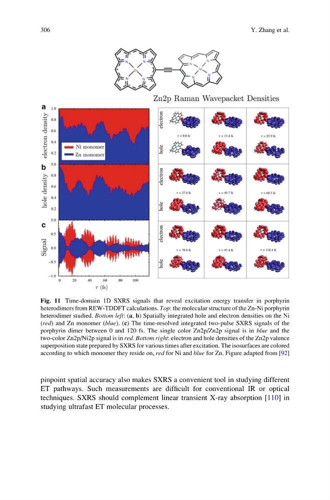
Fig. 11 Time-domain 1D SXRS signals that reveal excitation energy transfer in porphyrin
heterodimers from REW-TDDFT calculations. Top: the molecular structure of the Zn-Ni porphyrin
heterodimer studied. Bottom left: (a, b) Spatially integrated hole and electron densities on the Ni
(red) and Zn monomer (blue). (c) The time-resolved integrated two-pulse SXRS signals of the
porphyrin dimer between 0 and 120 fs. The single color Zn2p/Zn2p signal is in blue and the
two-color Zn2p/Ni2p signal is in red. Bottom right: electron and hole densities of the Zn2p valence
superposition state prepared by SXRS for various times after excitation. The isosurfaces are colored
according to which monomer they reside on, red for Ni and blue for Zn. Figure adapted from [92]
306
Y. Zhang et al.
ET pathways. Such measurements are difficult for conventional IR or optical
techniques. SXRS should complement linear transient X-ray absorption [110] in
studying ultrafast ET molecular processes.
Fig. 11 Time-domain 1D SXRS signals that reveal excitation energy transfer in porphyrin
heterodimers from REW-TDDFT calculations. Top: the molecular structure of the Zn-Ni porphyrin
heterodimer studied. Bottom left: (a, b) Spatially integrated hole and electron densities on the Ni
(red) and Zn monomer (blue). (c) The time-resolved integrated two-pulse SXRS signals of the
porphyrin dimer between 0 and 120 fs. The single color Zn2p/Zn2p signal is in blue and the
two-color Zn2p/Ni2p signal is in red. Bottom right: electron and hole densities of the Zn2p valence
superposition state prepared by SXRS for various times after excitation. The isosurfaces are colored
according to which monomer they reside on, red for Ni and blue for Zn. Figure adapted from [92]
306
Y. Zhang et al.