

Correlation between the motion of the doorway wavepacket and the fluctuations of
the SXRS signal profiles can be established. The time-domain signals provide a
real-time image of EET in the system, which is not possible for time-resolved
fluorescence anisotropy decay studies. SXRS could become a powerful tool in
revealing EET mechanisms in molecular systems. Further geometrical and structural factors that control EET in a series of porphyrin heterodimer systems were
studied in [93].
In another SXRS simulation study we investigated long-range electron transfer
(ET) in the small redox protein azurin [109]. Borrowing the ET kinetic parameters
from time-resolved infrared (IR) and optical measurements, time-resolved SXRS
signals at the electron donor, hopping intermediate and electron acceptor were
simulated with REW-TDDFT. We found that the SXRS signals depend sensitively
on the local electronic structure changes around the excited atoms, and could serve
as an excellent indicator for detecting electron transfer dynamics. The atomic
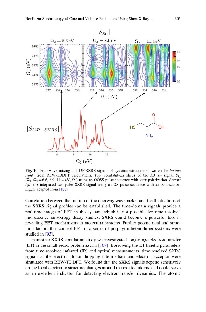
Fig. 10 Four-wave mixing and I2P-SXRS signals of cysteine (structure shown on the bottom
right) from REW-TDDFT calculations. Top: constatnt-Ω 2 slices of the 3D k II signal S k II
(Ω 1 , Ω 2 ¼ 6.6, 8.9, 11.4 eV, Ω 3 ) using an OOSS pulse sequence with xxxx polarization. Bottom
left: the integrated two-pulse SXRS signal using an OS pulse sequence with xx polarization.
Figure adapted from [108]
Nonlinear Spectroscopy of Core and Valence Excitations Using Short X-Ray. . .
305
the SXRS signal profiles can be established. The time-domain signals provide a
real-time image of EET in the system, which is not possible for time-resolved
fluorescence anisotropy decay studies. SXRS could become a powerful tool in
revealing EET mechanisms in molecular systems. Further geometrical and structural factors that control EET in a series of porphyrin heterodimer systems were
studied in [93].
In another SXRS simulation study we investigated long-range electron transfer
(ET) in the small redox protein azurin [109]. Borrowing the ET kinetic parameters
from time-resolved infrared (IR) and optical measurements, time-resolved SXRS
signals at the electron donor, hopping intermediate and electron acceptor were
simulated with REW-TDDFT. We found that the SXRS signals depend sensitively
on the local electronic structure changes around the excited atoms, and could serve
as an excellent indicator for detecting electron transfer dynamics. The atomic
Fig. 10 Four-wave mixing and I2P-SXRS signals of cysteine (structure shown on the bottom
right) from REW-TDDFT calculations. Top: constatnt-Ω 2 slices of the 3D k II signal S k II
(Ω 1 , Ω 2 ¼ 6.6, 8.9, 11.4 eV, Ω 3 ) using an OOSS pulse sequence with xxxx polarization. Bottom
left: the integrated two-pulse SXRS signal using an OS pulse sequence with xx polarization.
Figure adapted from [108]
Nonlinear Spectroscopy of Core and Valence Excitations Using Short X-Ray. . .
305