

relative flexibility that characterizes each of these components. In this study such
analysis was performed for each amino acid residue, considering the C α carbon
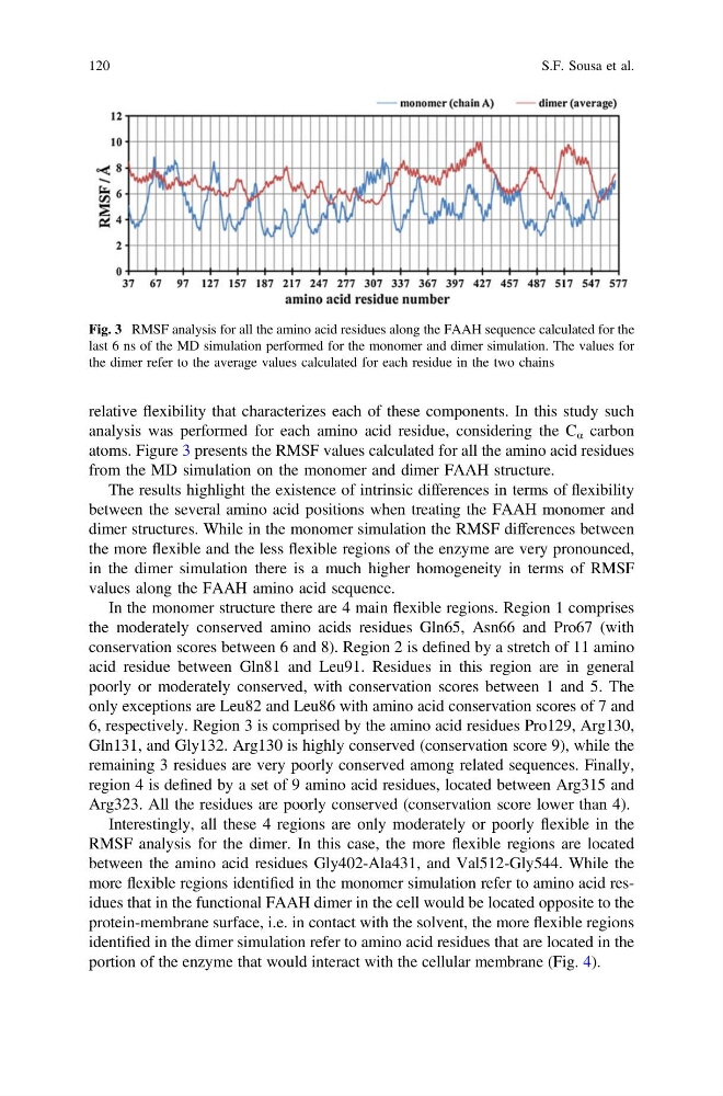
atoms. Figure 3 presents the RMSF values calculated for all the amino acid residues
from the MD simulation on the monomer and dimer FAAH structure.
The results highlight the existence of intrinsic differences in terms of flexibility
between the several amino acid positions when treating the FAAH monomer and
dimer structures. While in the monomer simulation the RMSF differences between
the more flexible and the less flexible regions of the enzyme are very pronounced,
in the dimer simulation there is a much higher homogeneity in terms of RMSF
values along the FAAH amino acid sequence.
In the monomer structure there are 4 main flexible regions. Region 1 comprises
the moderately conserved amino acids residues Gln65, Asn66 and Pro67 (with
conservation scores between 6 and 8). Region 2 is defined by a stretch of 11 amino
acid residue between Gln81 and Leu91. Residues in this region are in general
poorly or moderately conserved, with conservation scores between 1 and 5. The
only exceptions are Leu82 and Leu86 with amino acid conservation scores of 7 and
6, respectively. Region 3 is comprised by the amino acid residues Pro129, Arg130,
Gln131, and Gly132. Arg130 is highly conserved (conservation score 9), while the
remaining 3 residues are very poorly conserved among related sequences. Finally,
region 4 is defined by a set of 9 amino acid residues, located between Arg315 and
Arg323. All the residues are poorly conserved (conservation score lower than 4).
Interestingly, all these 4 regions are only moderately or poorly flexible in the
RMSF analysis for the dimer. In this case, the more flexible regions are located
between the amino acid residues Gly402-Ala431, and Val512-Gly544. While the
more flexible regions identified in the monomer simulation refer to amino acid residues that in the functional FAAH dimer in the cell would be located opposite to the
protein-membrane surface, i.e. in contact with the solvent, the more flexible regions
identified in the dimer simulation refer to amino acid residues that are located in the
portion of the enzyme that would interact with the cellular membrane (Fig. 4).
Fig. 3 RMSF analysis for all the amino acid residues along the FAAH sequence calculated for the
last 6 ns of the MD simulation performed for the monomer and dimer simulation. The values for
the dimer refer to the average values calculated for each residue in the two chains
120
S.F. Sousa et al.
analysis was performed for each amino acid residue, considering the C α carbon
atoms. Figure 3 presents the RMSF values calculated for all the amino acid residues
from the MD simulation on the monomer and dimer FAAH structure.
The results highlight the existence of intrinsic differences in terms of flexibility
between the several amino acid positions when treating the FAAH monomer and
dimer structures. While in the monomer simulation the RMSF differences between
the more flexible and the less flexible regions of the enzyme are very pronounced,
in the dimer simulation there is a much higher homogeneity in terms of RMSF
values along the FAAH amino acid sequence.
In the monomer structure there are 4 main flexible regions. Region 1 comprises
the moderately conserved amino acids residues Gln65, Asn66 and Pro67 (with
conservation scores between 6 and 8). Region 2 is defined by a stretch of 11 amino
acid residue between Gln81 and Leu91. Residues in this region are in general
poorly or moderately conserved, with conservation scores between 1 and 5. The
only exceptions are Leu82 and Leu86 with amino acid conservation scores of 7 and
6, respectively. Region 3 is comprised by the amino acid residues Pro129, Arg130,
Gln131, and Gly132. Arg130 is highly conserved (conservation score 9), while the
remaining 3 residues are very poorly conserved among related sequences. Finally,
region 4 is defined by a set of 9 amino acid residues, located between Arg315 and
Arg323. All the residues are poorly conserved (conservation score lower than 4).
Interestingly, all these 4 regions are only moderately or poorly flexible in the
RMSF analysis for the dimer. In this case, the more flexible regions are located
between the amino acid residues Gly402-Ala431, and Val512-Gly544. While the
more flexible regions identified in the monomer simulation refer to amino acid residues that in the functional FAAH dimer in the cell would be located opposite to the
protein-membrane surface, i.e. in contact with the solvent, the more flexible regions
identified in the dimer simulation refer to amino acid residues that are located in the
portion of the enzyme that would interact with the cellular membrane (Fig. 4).
Fig. 3 RMSF analysis for all the amino acid residues along the FAAH sequence calculated for the
last 6 ns of the MD simulation performed for the monomer and dimer simulation. The values for
the dimer refer to the average values calculated for each residue in the two chains
120
S.F. Sousa et al.