

Soluble elastin prepared by the method of Partridge [7] has an isoelectric point
around pH 5. Correspondingly, we measured isoelectric points between pH 5 and
6 for ES12-PEG-DGE and around 6 for BS3 crosslinked microgels. For the elastin
at low ionic strength, it has been reported that the coacervation temperatures exhibit
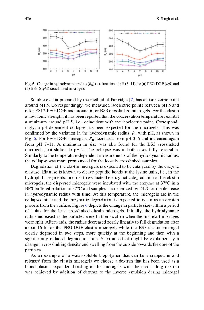
a minimum around pH 5, i.e., coincident with the isoelectric point. Correspondingly, a pH-dependent collapse has been expected for the microgels. This was
confirmed by the variation in the hydrodynamic radius, R h with pH, as shown in
Fig. 5. For PEG-DGE microgels, R h decreased from pH 3–6 and increased again
from pH 7–11. A minimum in size was also found for the BS3 crosslinked
microgels, but shifted to pH 7. The collapse was in both cases fully reversible.
Similarly to the temperature-dependent measurements of the hydrodynamic radius,
the collapse was more pronounced for the loosely crosslinked samples.
Degradation of the elastin microgels is expected to be catalyzed by the enzyme
elastase. Elastase is known to cleave peptidic bonds at the lysine units, i.e., in the
hydrophilic segments. In order to evaluate the enzymatic degradation of the elastin
microgels, the dispersed microgels were incubated with the enzyme at 37
C in a
BPS buffered solution at 37
C and samples characterized by DLS for the decrease
in hydrodynamic radius with time. At this temperature, the microgels are in the
collapsed state and the enzymatic degradation is expected to occur as an erosion
process from the surface. Figure 6 depicts the change in particle size within a period
of 1 day for the least crosslinked elastin microgels. Initially, the hydrodynamic
radius increased as the particles were further swollen when the first elastin bridges
were split. Afterwards, the radius decreased nearly linearly to full degradation after
about 16 h for the PEG-DGE-elastin microgel, while the BS3-elastin microgel
clearly degraded in two steps, more quickly at the beginning and then with a
significantly reduced degradation rate. Such an effect might be explained by a
change in crosslinking density and swelling from the outside towards the core of the
particles.
As an example of a water-soluble biopolymer that can be entrapped in and
released from the elastin microgels we choose a dextran that has been used as a
blood plasma expander. Loading of the microgels with the model drug dextran
was achieved by addition of dextran to the inverse emulsion during microgel
Fig. 5 Change in hydrodynamic radius (R h ) as a function of pH (3–11) for (a) PEG-DGE (left) and
(b) BS3 (right) crosslinked microgels
426
S. Singh et al.
around pH 5. Correspondingly, we measured isoelectric points between pH 5 and
6 for ES12-PEG-DGE and around 6 for BS3 crosslinked microgels. For the elastin
at low ionic strength, it has been reported that the coacervation temperatures exhibit
a minimum around pH 5, i.e., coincident with the isoelectric point. Correspondingly, a pH-dependent collapse has been expected for the microgels. This was
confirmed by the variation in the hydrodynamic radius, R h with pH, as shown in
Fig. 5. For PEG-DGE microgels, R h decreased from pH 3–6 and increased again
from pH 7–11. A minimum in size was also found for the BS3 crosslinked
microgels, but shifted to pH 7. The collapse was in both cases fully reversible.
Similarly to the temperature-dependent measurements of the hydrodynamic radius,
the collapse was more pronounced for the loosely crosslinked samples.
Degradation of the elastin microgels is expected to be catalyzed by the enzyme
elastase. Elastase is known to cleave peptidic bonds at the lysine units, i.e., in the
hydrophilic segments. In order to evaluate the enzymatic degradation of the elastin
microgels, the dispersed microgels were incubated with the enzyme at 37
C in a
BPS buffered solution at 37
C and samples characterized by DLS for the decrease
in hydrodynamic radius with time. At this temperature, the microgels are in the
collapsed state and the enzymatic degradation is expected to occur as an erosion
process from the surface. Figure 6 depicts the change in particle size within a period
of 1 day for the least crosslinked elastin microgels. Initially, the hydrodynamic
radius increased as the particles were further swollen when the first elastin bridges
were split. Afterwards, the radius decreased nearly linearly to full degradation after
about 16 h for the PEG-DGE-elastin microgel, while the BS3-elastin microgel
clearly degraded in two steps, more quickly at the beginning and then with a
significantly reduced degradation rate. Such an effect might be explained by a
change in crosslinking density and swelling from the outside towards the core of the
particles.
As an example of a water-soluble biopolymer that can be entrapped in and
released from the elastin microgels we choose a dextran that has been used as a
blood plasma expander. Loading of the microgels with the model drug dextran
was achieved by addition of dextran to the inverse emulsion during microgel
Fig. 5 Change in hydrodynamic radius (R h ) as a function of pH (3–11) for (a) PEG-DGE (left) and
(b) BS3 (right) crosslinked microgels
426
S. Singh et al.