

H-bonding. Therefore, it can be viewed as a starting point for the creation of loops
that undertake relevant structural changes upon binding to an external partner
(solvent, another peptide, etc.). This is, after all, an important feature of loops in
proteins [82].
Shortly after this first study, Gorske and Blackwell were able to show that
incorporation of fluorinated aromatics modified the folding behavior. Incorporation
of only one unit of 1-pentafluorophenylethyl residue at the N-terminus of a nonamer
stabilized the loop. As a result, addition of a protic solvent (methanol) was better
tolerated with respect to the conservation of the loop structure [84].
Later, it was shown that other small structural changes within one monomer unit
can have a strong influence on the formation of secondary structures. Blackwell
et al. prepared peptoid octamers of the helicogenic Nspe that carried a single
1-(nitrophenyl)ethyl residue at the N-terminus. Interestingly, the position of the
nitro-moiety (2 or 4 of the phenyl ring) either destabilized or stabilized the threaded
loop discussed previously for Nspe 9 [85].
Two different approaches were used to induce formation of turns, another
important structural motif in proteins. Both approaches have in common that it
was necessary to deviate from the peptoid backbone [25, 86]. Apella and coworkers
employed a triazole ring to induce a hairpin-like structure (Fig. 9a) [86]. Importantly, this secondary structure was confirmed in aqueous media. In contrast,
Blackwell and colleagues introduced a tripeptoid, in which one monomer unit
was attached to a peptoid side chain. Moreover, a critical design element was that
this substituent was able to undergo H-bonding (Fig. 9b) [25]. Thus, it may be
argued that the unique character of peptoid-based secondary structures, which
typically are ensured without employment of H-bonding, is lost to some degree.
Very recently, Blackwell and coworkers reported on a peptoid-based ribbon
structure. Such ribbons are also found in proteins and may function as
cell-membrane-modifying agents and antibiotics [87]. The ribbon structure was
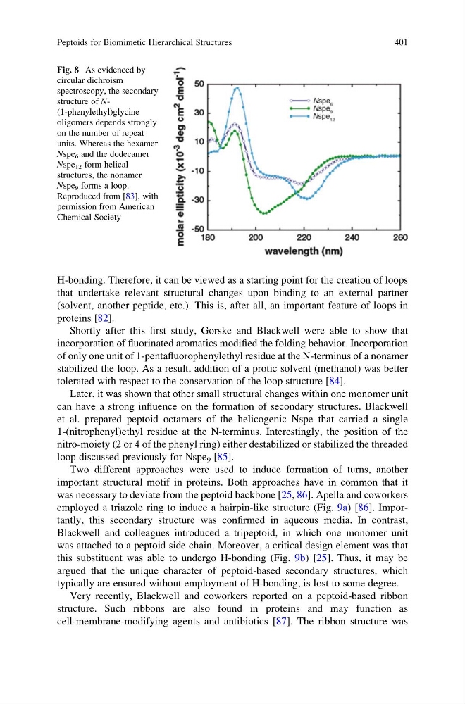
Fig. 8 As evidenced by
circular dichroism
spectroscopy, the secondary
structure of N(1-phenylethyl)glycine
oligomers depends strongly
on the number of repeat
units. Whereas the hexamer
Nspe 6 and the dodecamer
Nspe 12 form helical
structures, the nonamer
Nspe 9 forms a loop.
Reproduced from [83], with
permission from American
Chemical Society
Peptoids for Biomimetic Hierarchical Structures
401
that undertake relevant structural changes upon binding to an external partner
(solvent, another peptide, etc.). This is, after all, an important feature of loops in
proteins [82].
Shortly after this first study, Gorske and Blackwell were able to show that
incorporation of fluorinated aromatics modified the folding behavior. Incorporation
of only one unit of 1-pentafluorophenylethyl residue at the N-terminus of a nonamer
stabilized the loop. As a result, addition of a protic solvent (methanol) was better
tolerated with respect to the conservation of the loop structure [84].
Later, it was shown that other small structural changes within one monomer unit
can have a strong influence on the formation of secondary structures. Blackwell
et al. prepared peptoid octamers of the helicogenic Nspe that carried a single
1-(nitrophenyl)ethyl residue at the N-terminus. Interestingly, the position of the
nitro-moiety (2 or 4 of the phenyl ring) either destabilized or stabilized the threaded
loop discussed previously for Nspe 9 [85].
Two different approaches were used to induce formation of turns, another
important structural motif in proteins. Both approaches have in common that it
was necessary to deviate from the peptoid backbone [25, 86]. Apella and coworkers
employed a triazole ring to induce a hairpin-like structure (Fig. 9a) [86]. Importantly, this secondary structure was confirmed in aqueous media. In contrast,
Blackwell and colleagues introduced a tripeptoid, in which one monomer unit
was attached to a peptoid side chain. Moreover, a critical design element was that
this substituent was able to undergo H-bonding (Fig. 9b) [25]. Thus, it may be
argued that the unique character of peptoid-based secondary structures, which
typically are ensured without employment of H-bonding, is lost to some degree.
Very recently, Blackwell and coworkers reported on a peptoid-based ribbon
structure. Such ribbons are also found in proteins and may function as
cell-membrane-modifying agents and antibiotics [87]. The ribbon structure was
Fig. 8 As evidenced by
circular dichroism
spectroscopy, the secondary
structure of N(1-phenylethyl)glycine
oligomers depends strongly
on the number of repeat
units. Whereas the hexamer
Nspe 6 and the dodecamer
Nspe 12 form helical
structures, the nonamer
Nspe 9 forms a loop.
Reproduced from [83], with
permission from American
Chemical Society
Peptoids for Biomimetic Hierarchical Structures
401