

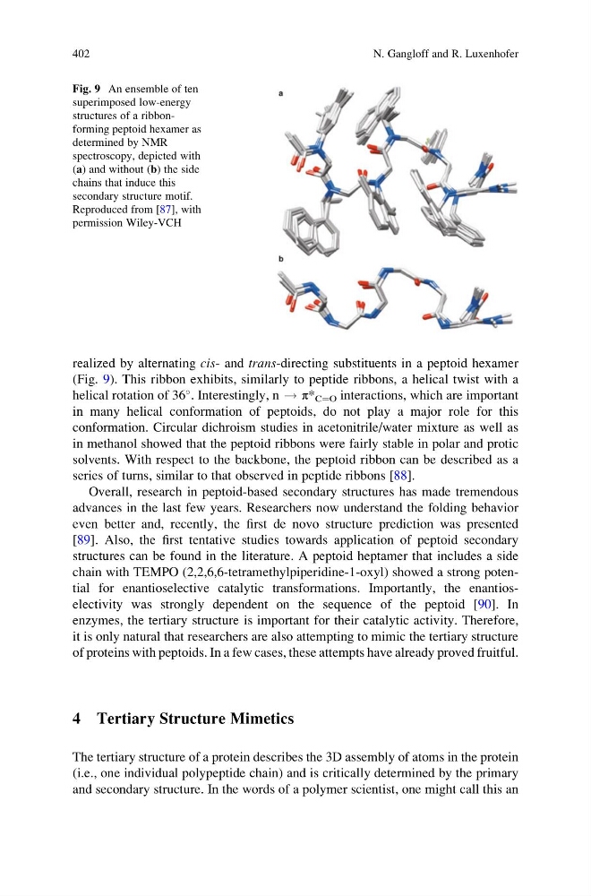
realized by alternating cis- and trans-directing substituents in a peptoid hexamer
(Fig. 9). This ribbon exhibits, similarly to peptide ribbons, a helical twist with a
helical rotation of 36
. Interestingly, n ! π* C¼O interactions, which are important
in many helical conformation of peptoids, do not play a major role for this
conformation. Circular dichroism studies in acetonitrile/water mixture as well as
in methanol showed that the peptoid ribbons were fairly stable in polar and protic
solvents. With respect to the backbone, the peptoid ribbon can be described as a
series of turns, similar to that observed in peptide ribbons [88].
Overall, research in peptoid-based secondary structures has made tremendous
advances in the last few years. Researchers now understand the folding behavior
even better and, recently, the first de novo structure prediction was presented
[89]. Also, the first tentative studies towards application of peptoid secondary
structures can be found in the literature. A peptoid heptamer that includes a side
chain with TEMPO (2,2,6,6-tetramethylpiperidine-1-oxyl) showed a strong potential for enantioselective catalytic transformations. Importantly, the enantioselectivity was strongly dependent on the sequence of the peptoid [90]. In
enzymes, the tertiary structure is important for their catalytic activity. Therefore,
it is only natural that researchers are also attempting to mimic the tertiary structure
of proteins with peptoids. In a few cases, these attempts have already proved fruitful.
4 Tertiary Structure Mimetics
The tertiary structure of a protein describes the 3D assembly of atoms in the protein
(i.e., one individual polypeptide chain) and is critically determined by the primary
and secondary structure. In the words of a polymer scientist, one might call this an
Fig. 9 An ensemble of ten
superimposed low-energy
structures of a ribbonforming peptoid hexamer as
determined by NMR
spectroscopy, depicted with
(a) and without (b) the side
chains that induce this
secondary structure motif.
Reproduced from [87], with
permission Wiley-VCH
402
N. Gangloff and R. Luxenhofer
(Fig. 9). This ribbon exhibits, similarly to peptide ribbons, a helical twist with a
helical rotation of 36
. Interestingly, n ! π* C¼O interactions, which are important
in many helical conformation of peptoids, do not play a major role for this
conformation. Circular dichroism studies in acetonitrile/water mixture as well as
in methanol showed that the peptoid ribbons were fairly stable in polar and protic
solvents. With respect to the backbone, the peptoid ribbon can be described as a
series of turns, similar to that observed in peptide ribbons [88].
Overall, research in peptoid-based secondary structures has made tremendous
advances in the last few years. Researchers now understand the folding behavior
even better and, recently, the first de novo structure prediction was presented
[89]. Also, the first tentative studies towards application of peptoid secondary
structures can be found in the literature. A peptoid heptamer that includes a side
chain with TEMPO (2,2,6,6-tetramethylpiperidine-1-oxyl) showed a strong potential for enantioselective catalytic transformations. Importantly, the enantioselectivity was strongly dependent on the sequence of the peptoid [90]. In
enzymes, the tertiary structure is important for their catalytic activity. Therefore,
it is only natural that researchers are also attempting to mimic the tertiary structure
of proteins with peptoids. In a few cases, these attempts have already proved fruitful.
4 Tertiary Structure Mimetics
The tertiary structure of a protein describes the 3D assembly of atoms in the protein
(i.e., one individual polypeptide chain) and is critically determined by the primary
and secondary structure. In the words of a polymer scientist, one might call this an
Fig. 9 An ensemble of ten
superimposed low-energy
structures of a ribbonforming peptoid hexamer as
determined by NMR
spectroscopy, depicted with
(a) and without (b) the side
chains that induce this
secondary structure motif.
Reproduced from [87], with
permission Wiley-VCH
402
N. Gangloff and R. Luxenhofer