

(and to some extent, the innermost vesicle) also experience a strongly anisotropic
environment due to the fact that one of the leaflets is within a multilamellar environment and the other faces the bulk water solution. Obviously, one expects that bulk
mechanical properties are influenced by such anisotropy. Although a multinanometer
thick layer of water separates two adjacent vesicles, such water phase is highly
incompressible and has the effect of linking the deformations of the contained vesicle
to those of the containing one. However, the multilamellar arrangement can also have
an effect on the “microscopic” properties, such as the diffusion times of individual
lipid molecules. This hypothesis can be tested easily by CG–MD simulations with
explicit solvent.
We prepared ULVs made by 1,512, 2,500, 3,500, and 5,000 DMPC
(1,2-dimyristoyl-sn-glycero-3-phosphocholine) lipid molecules, respectively. These
vesicles were prepared by a series of MD simulations. Briefly, the prepared vesicles
are mostly stress-free, because those structures are spontaneously formed from an
arbitrary initial aggregate structure during 100–200 ns CG–MD. Timing is supposed
to be long enough to see a reasonable partitioning of lipids between inner and outer
leaflets of the membrane to relax the stress. We also carried out MD simulation of an
MLV, generated by a combination of two ULVs containing 1,512 and 5,000 DMPC
lipids, respectively; we simply placed the smaller vesicle inside the larger vesicle.
A flat membrane made by 8,194 DMPC lipid molecules was also investigated using
three-dimensional periodic boundary conditions, which effectively mimic the
multilamellar stack phase.
MD simulations were carried out in the NPT ensemble, with the temperature at
310 K and pressure set at 1 atm. MD simulation of each vesicle system was
conducted for 1 μs, although the MD run of the flat membrane was performed for
only 300 ns.
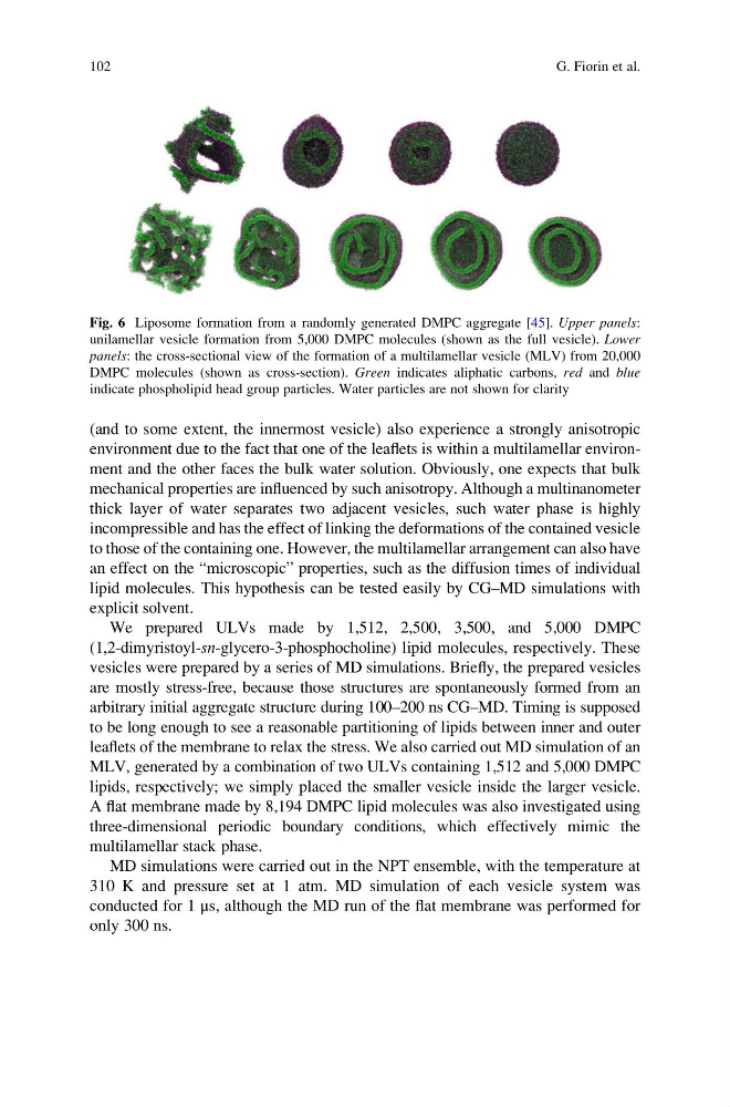
Fig. 6 Liposome formation from a randomly generated DMPC aggregate [45]. Upper panels:
unilamellar vesicle formation from 5,000 DMPC molecules (shown as the full vesicle). Lower
panels: the cross-sectional view of the formation of a multilamellar vesicle (MLV) from 20,000
DMPC molecules (shown as cross-section). Green indicates aliphatic carbons, red and blue
indicate phospholipid head group particles. Water particles are not shown for clarity
102
G. Fiorin et al.
environment due to the fact that one of the leaflets is within a multilamellar environment and the other faces the bulk water solution. Obviously, one expects that bulk
mechanical properties are influenced by such anisotropy. Although a multinanometer
thick layer of water separates two adjacent vesicles, such water phase is highly
incompressible and has the effect of linking the deformations of the contained vesicle
to those of the containing one. However, the multilamellar arrangement can also have
an effect on the “microscopic” properties, such as the diffusion times of individual
lipid molecules. This hypothesis can be tested easily by CG–MD simulations with
explicit solvent.
We prepared ULVs made by 1,512, 2,500, 3,500, and 5,000 DMPC
(1,2-dimyristoyl-sn-glycero-3-phosphocholine) lipid molecules, respectively. These
vesicles were prepared by a series of MD simulations. Briefly, the prepared vesicles
are mostly stress-free, because those structures are spontaneously formed from an
arbitrary initial aggregate structure during 100–200 ns CG–MD. Timing is supposed
to be long enough to see a reasonable partitioning of lipids between inner and outer
leaflets of the membrane to relax the stress. We also carried out MD simulation of an
MLV, generated by a combination of two ULVs containing 1,512 and 5,000 DMPC
lipids, respectively; we simply placed the smaller vesicle inside the larger vesicle.
A flat membrane made by 8,194 DMPC lipid molecules was also investigated using
three-dimensional periodic boundary conditions, which effectively mimic the
multilamellar stack phase.
MD simulations were carried out in the NPT ensemble, with the temperature at
310 K and pressure set at 1 atm. MD simulation of each vesicle system was
conducted for 1 μs, although the MD run of the flat membrane was performed for
only 300 ns.
Fig. 6 Liposome formation from a randomly generated DMPC aggregate [45]. Upper panels:
unilamellar vesicle formation from 5,000 DMPC molecules (shown as the full vesicle). Lower
panels: the cross-sectional view of the formation of a multilamellar vesicle (MLV) from 20,000
DMPC molecules (shown as cross-section). Green indicates aliphatic carbons, red and blue
indicate phospholipid head group particles. Water particles are not shown for clarity
102
G. Fiorin et al.