

6 Assembly of Multilamellar Vesicles
Two-dimensional membranes may easily be replicated in three dimensions to produce
entirely new morphologies. The simplest example is the multilamellar stack phase,
observed at low humidity (less than 50%). After that, a more interesting system is the
multilamellar vesicle (MLV), where each unilamellar vesicle (ULV), the containing
one and the contained one, has different properties (Fig. 6). The outermost vesicle
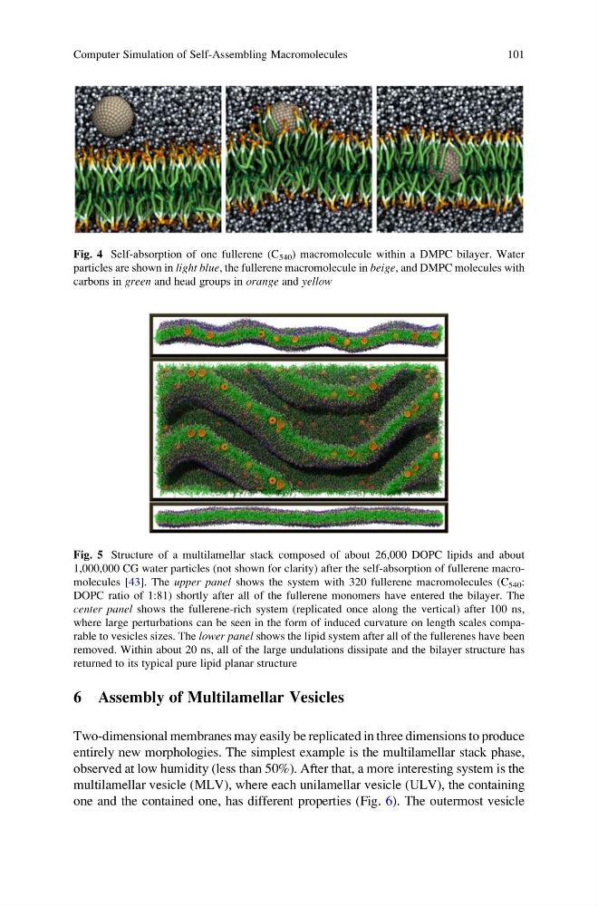
Fig. 4 Self-absorption of one fullerene (C 540 ) macromolecule within a DMPC bilayer. Water
particles are shown in light blue, the fullerene macromolecule in beige, and DMPC molecules with
carbons in green and head groups in orange and yellow
Fig. 5 Structure of a multilamellar stack composed of about 26,000 DOPC lipids and about
1,000,000 CG water particles (not shown for clarity) after the self-absorption of fullerene macromolecules [43]. The upper panel shows the system with 320 fullerene macromolecules (C 540 :
DOPC ratio of 1:81) shortly after all of the fullerene monomers have entered the bilayer. The
center panel shows the fullerene-rich system (replicated once along the vertical) after 100 ns,
where large perturbations can be seen in the form of induced curvature on length scales comparable to vesicles sizes. The lower panel shows the lipid system after all of the fullerenes have been
removed. Within about 20 ns, all of the large undulations dissipate and the bilayer structure has
returned to its typical pure lipid planar structure
Computer Simulation of Self-Assembling Macromolecules
101
Two-dimensional membranes may easily be replicated in three dimensions to produce
entirely new morphologies. The simplest example is the multilamellar stack phase,
observed at low humidity (less than 50%). After that, a more interesting system is the
multilamellar vesicle (MLV), where each unilamellar vesicle (ULV), the containing
one and the contained one, has different properties (Fig. 6). The outermost vesicle
Fig. 4 Self-absorption of one fullerene (C 540 ) macromolecule within a DMPC bilayer. Water
particles are shown in light blue, the fullerene macromolecule in beige, and DMPC molecules with
carbons in green and head groups in orange and yellow
Fig. 5 Structure of a multilamellar stack composed of about 26,000 DOPC lipids and about
1,000,000 CG water particles (not shown for clarity) after the self-absorption of fullerene macromolecules [43]. The upper panel shows the system with 320 fullerene macromolecules (C 540 :
DOPC ratio of 1:81) shortly after all of the fullerene monomers have entered the bilayer. The
center panel shows the fullerene-rich system (replicated once along the vertical) after 100 ns,
where large perturbations can be seen in the form of induced curvature on length scales comparable to vesicles sizes. The lower panel shows the lipid system after all of the fullerenes have been
removed. Within about 20 ns, all of the large undulations dissipate and the bilayer structure has
returned to its typical pure lipid planar structure
Computer Simulation of Self-Assembling Macromolecules
101