

its components. As early as the beginning of the twentieth century, the composition of
biological membranes had correctly been understood as a mixture of phospholipid and
cholesterol by Meyer and Overton, during their studies of the mechanisms of anesthesia [46]. However, it was not until decades later that the bilayer structure could be
inferred [47], and not until the mid-twentieth century that this structure could be
observed directly by electron microscopy [48, 49].
Only recently have direct MD simulations methods been able to observe the selfassembly of a phospholipid bilayer. An ab initio description of the macromolecules
involved that includes its electrons is of course impractical due to its computational
cost. However, even approaches where the potential energy function is an empirical
force field [15–19] have proven insufficient to observe the self-assembly of a small
membrane. For a few hundreds of phospholipid molecules to assemble in a bilayer a
few nanometers wide, time scales well beyond the microsecond are required.
Although the accessible times by MD simulations have increased dramatically,
only in recent years has the microsecond threshold been surpassed effectively.
The availability of CG models for phospholipids has made it possible to observe
the self-assembly of Langmuir monolayers of phospholipids [31], bilayers of
nonionic surfactants [38], and finally of phospholipids [41], all beginning from
initial conditions where lipid or surfactant molecules are completely dissolved in
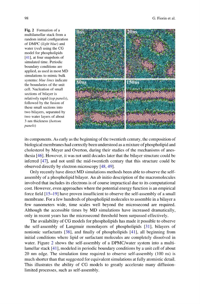
water. Figure 2 shows the self-assembly of a DPMC/water system into a multilamellar stack [41], modeled in periodic boundary conditions by a unit cell of about
20 nm edge. The simulation time required to observe self-assembly (100 ns) is
much shorter than that suggested for equivalent simulations at fully atomistic detail.
This illustrates the ability of CG models to greatly accelerate many diffusionlimited processes, such as self-assembly.
Fig. 2 Formation of a
multilamellar stack from a
random initial configuration
of DMPC (light blue) and
water (red) using the CG
model for phospholipids
[41], at four snapshots of
simulated time. Periodic
boundary conditions are
applied, as used in most MD
simulations to mimic bulk
systems: blue lines indicate
the boundaries of the unit
cell. Nucleation of small
sections of bilayer is
relatively rapid (top panels),
followed by the fusion of
these small sections into
two bilayers, separated by
two water layers of about
3 nm thickness (bottom
panels)
98
G. Fiorin et al.
biological membranes had correctly been understood as a mixture of phospholipid and
cholesterol by Meyer and Overton, during their studies of the mechanisms of anesthesia [46]. However, it was not until decades later that the bilayer structure could be
inferred [47], and not until the mid-twentieth century that this structure could be
observed directly by electron microscopy [48, 49].
Only recently have direct MD simulations methods been able to observe the selfassembly of a phospholipid bilayer. An ab initio description of the macromolecules
involved that includes its electrons is of course impractical due to its computational
cost. However, even approaches where the potential energy function is an empirical
force field [15–19] have proven insufficient to observe the self-assembly of a small
membrane. For a few hundreds of phospholipid molecules to assemble in a bilayer a
few nanometers wide, time scales well beyond the microsecond are required.
Although the accessible times by MD simulations have increased dramatically,
only in recent years has the microsecond threshold been surpassed effectively.
The availability of CG models for phospholipids has made it possible to observe
the self-assembly of Langmuir monolayers of phospholipids [31], bilayers of
nonionic surfactants [38], and finally of phospholipids [41], all beginning from
initial conditions where lipid or surfactant molecules are completely dissolved in
water. Figure 2 shows the self-assembly of a DPMC/water system into a multilamellar stack [41], modeled in periodic boundary conditions by a unit cell of about
20 nm edge. The simulation time required to observe self-assembly (100 ns) is
much shorter than that suggested for equivalent simulations at fully atomistic detail.
This illustrates the ability of CG models to greatly accelerate many diffusionlimited processes, such as self-assembly.
Fig. 2 Formation of a
multilamellar stack from a
random initial configuration
of DMPC (light blue) and
water (red) using the CG
model for phospholipids
[41], at four snapshots of
simulated time. Periodic
boundary conditions are
applied, as used in most MD
simulations to mimic bulk
systems: blue lines indicate
the boundaries of the unit
cell. Nucleation of small
sections of bilayer is
relatively rapid (top panels),
followed by the fusion of
these small sections into
two bilayers, separated by
two water layers of about
3 nm thickness (bottom
panels)
98
G. Fiorin et al.