

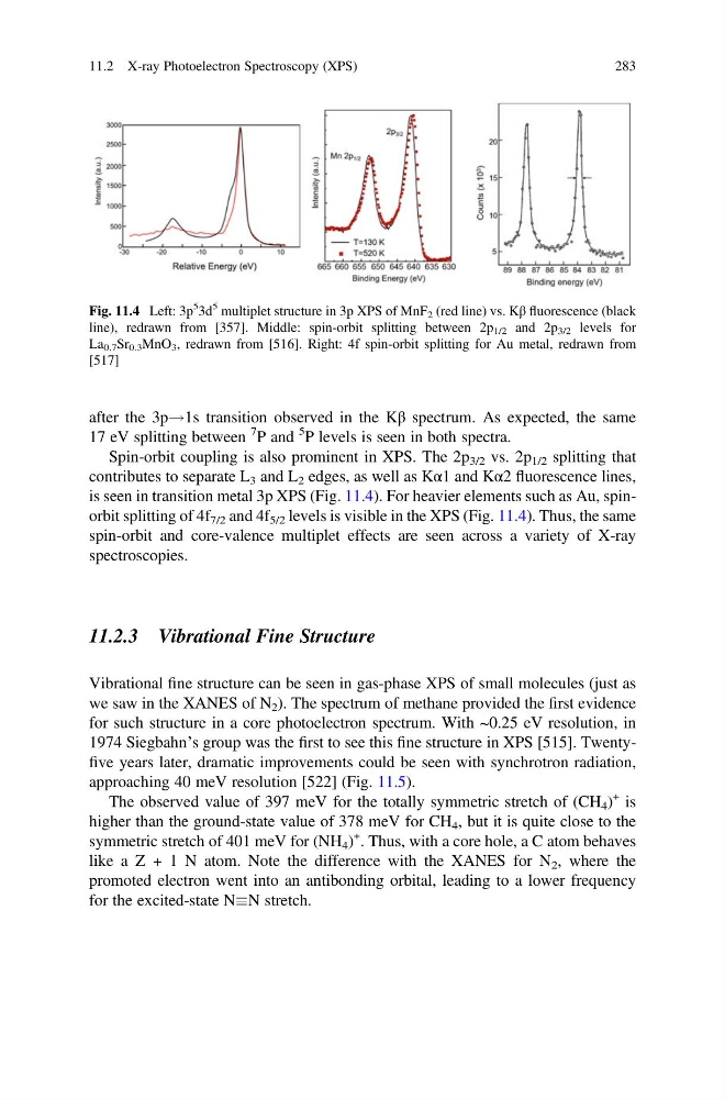
after the 3p!1s transition observed in the Kβ spectrum. As expected, the same
17 eV splitting between
7 P and
5 P levels is seen in both spectra.
Spin-orbit coupling is also prominent in XPS. The 2p 3/2 vs. 2p 1/2 splitting that
contributes to separate L 3 and L 2 edges, as well as Kα1 and Kα2 fluorescence lines,
is seen in transition metal 3p XPS (Fig. 11.4). For heavier elements such as Au, spinorbit splitting of 4f 7/2 and 4f 5/2 levels is visible in the XPS (Fig. 11.4). Thus, the same
spin-orbit and core-valence multiplet effects are seen across a variety of X-ray
spectroscopies.
11.2.3 Vibrational Fine Structure
Vibrational fine structure can be seen in gas-phase XPS of small molecules (just as
we saw in the XANES of N 2 ). The spectrum of methane provided the first evidence
for such structure in a core photoelectron spectrum. With ~0.25 eV resolution, in
1974 Siegbahn’s group was the first to see this fine structure in XPS [515]. Twentyfive years later, dramatic improvements could be seen with synchrotron radiation,
approaching 40 meV resolution [522] (Fig. 11.5).
The observed value of 397 meV for the totally symmetric stretch of (CH 4 )
+ is
higher than the ground-state value of 378 meV for CH 4 , but it is quite close to the
symmetric stretch of 401 meV for (NH 4 )
+ . Thus, with a core hole, a C atom behaves
like a Z + 1 N atom. Note the difference with the XANES for N 2 , where the
promoted electron went into an antibonding orbital, leading to a lower frequency
for the excited-state NN stretch.
Fig. 11.4 Left: 3p
5
3d
5 multiplet structure in 3p XPS of MnF 2 (red line) vs. Kβ fluorescence (black
line), redrawn from [357]. Middle: spin-orbit splitting between 2p 1/2 and 2p 3/2 levels for
La 0.7 Sr 0.3 MnO 3 , redrawn from [516]. Right: 4f spin-orbit splitting for Au metal, redrawn from
[517]
11.2 X-ray Photoelectron Spectroscopy (XPS)
283
17 eV splitting between
7 P and
5 P levels is seen in both spectra.
Spin-orbit coupling is also prominent in XPS. The 2p 3/2 vs. 2p 1/2 splitting that
contributes to separate L 3 and L 2 edges, as well as Kα1 and Kα2 fluorescence lines,
is seen in transition metal 3p XPS (Fig. 11.4). For heavier elements such as Au, spinorbit splitting of 4f 7/2 and 4f 5/2 levels is visible in the XPS (Fig. 11.4). Thus, the same
spin-orbit and core-valence multiplet effects are seen across a variety of X-ray
spectroscopies.
11.2.3 Vibrational Fine Structure
Vibrational fine structure can be seen in gas-phase XPS of small molecules (just as
we saw in the XANES of N 2 ). The spectrum of methane provided the first evidence
for such structure in a core photoelectron spectrum. With ~0.25 eV resolution, in
1974 Siegbahn’s group was the first to see this fine structure in XPS [515]. Twentyfive years later, dramatic improvements could be seen with synchrotron radiation,
approaching 40 meV resolution [522] (Fig. 11.5).
The observed value of 397 meV for the totally symmetric stretch of (CH 4 )
+ is
higher than the ground-state value of 378 meV for CH 4 , but it is quite close to the
symmetric stretch of 401 meV for (NH 4 )
+ . Thus, with a core hole, a C atom behaves
like a Z + 1 N atom. Note the difference with the XANES for N 2 , where the
promoted electron went into an antibonding orbital, leading to a lower frequency
for the excited-state NN stretch.
Fig. 11.4 Left: 3p
5
3d
5 multiplet structure in 3p XPS of MnF 2 (red line) vs. Kβ fluorescence (black
line), redrawn from [357]. Middle: spin-orbit splitting between 2p 1/2 and 2p 3/2 levels for
La 0.7 Sr 0.3 MnO 3 , redrawn from [516]. Right: 4f spin-orbit splitting for Au metal, redrawn from
[517]
11.2 X-ray Photoelectron Spectroscopy (XPS)
283