

asymmetrically cut crystal monochromator (Chap. 4). The output from the monochromator can then be refocused onto the sample with KB mirrors or other optics.
The sample, usually at cryogenic temperatures, may also be exposed to a high
magnetic field or other conditions. Finally, the scattered radiation is recorded with a
high-speed detector. Because it has excellent (<1 ns) time resolution, as well as the
ability to quickly recover from saturating overloads, the detector of choice has been
the avalanche photodiode [187,439] (Chap. 5). Both single elements and large arrays
[440] have been used.
9.5.2 The Refractive Index Model
There are a variety of approaches to describing nuclear forward scattering, including
the microscopic nuclear exciton model [441] and the macroscopic complex refractive index model [442]. Some of the mathematics for the latter approach are included
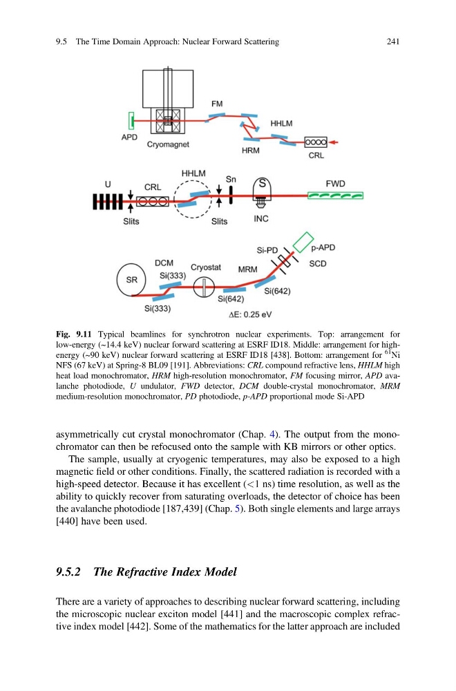
Fig. 9.11 Typical beamlines for synchrotron nuclear experiments. Top: arrangement for
low-energy (~14.4 keV) nuclear forward scattering at ESRF ID18. Middle: arrangement for highenergy (~90 keV) nuclear forward scattering at ESRF ID18 [438]. Bottom: arrangement for
61
Ni
NFS (67 keV) at Spring-8 BL09 [191]. Abbreviations: CRL compound refractive lens, HHLM high
heat load monochromator, HRM high-resolution monochromator, FM focusing mirror, APD avalanche photodiode, U undulator, FWD detector, DCM double-crystal monochromator, MRM
medium-resolution monochromator, PD photodiode, p-APD proportional mode Si-APD
9.5 The Time Domain Approach: Nuclear Forward Scattering
241
The sample, usually at cryogenic temperatures, may also be exposed to a high
magnetic field or other conditions. Finally, the scattered radiation is recorded with a
high-speed detector. Because it has excellent (<1 ns) time resolution, as well as the
ability to quickly recover from saturating overloads, the detector of choice has been
the avalanche photodiode [187,439] (Chap. 5). Both single elements and large arrays
[440] have been used.
9.5.2 The Refractive Index Model
There are a variety of approaches to describing nuclear forward scattering, including
the microscopic nuclear exciton model [441] and the macroscopic complex refractive index model [442]. Some of the mathematics for the latter approach are included
Fig. 9.11 Typical beamlines for synchrotron nuclear experiments. Top: arrangement for
low-energy (~14.4 keV) nuclear forward scattering at ESRF ID18. Middle: arrangement for highenergy (~90 keV) nuclear forward scattering at ESRF ID18 [438]. Bottom: arrangement for
61
Ni
NFS (67 keV) at Spring-8 BL09 [191]. Abbreviations: CRL compound refractive lens, HHLM high
heat load monochromator, HRM high-resolution monochromator, FM focusing mirror, APD avalanche photodiode, U undulator, FWD detector, DCM double-crystal monochromator, MRM
medium-resolution monochromator, PD photodiode, p-APD proportional mode Si-APD
9.5 The Time Domain Approach: Nuclear Forward Scattering
241