

scattering” or “NFS” experiment, the sample is excited by a very short synchrotron
pulse (<100 ps), and the forward-scattered intensity is monitored as a function of
time. The excitation is coherent because the sample is in the same state before and
after the scattering process. Since the particular atoms involved in the scattering
cannot be identified, one has to calculate the response as the sum over all possible
scattering paths.
9.5.1 NFS Methodology: The Synchrotron Experiment
Everything in a synchrotron nuclear experiment is designed to separate the small
nuclear signal from the overwhelming electronic background. Although peak
nuclear cross sections are large, they are also very narrow. For these experiments
to be successful, one requires (a) a high spectral brightness X-ray source (b) a very
high-resolution incident beam monochromator and (c) a high-speed detection system to separate nuclear events from ordinary X-ray scattering and fluorescence.
Typical arrangements are shown in Fig. 9.11.
Nowadays, the source is invariably an undulator on a third-generation synchrotron radiation facility or even an FEL. To reduce the vertical divergence, some
beamlines employ X-ray compound refractive lenses (Chap. 4). In either case, a high
heat load monochromator is used to select a narrow energy slice from the undulator
beam—otherwise the thermal load would overload the next stage, a high-resolution
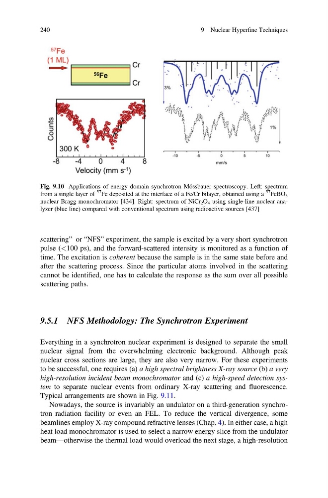
Fig. 9.10 Applications of energy domain synchrotron Mössbauer spectroscopy. Left: spectrum
from a single layer of
57
Fe deposited at the interface of a Fe/Cr bilayer, obtained using a
57
FeBO 3
nuclear Bragg monochromator [434]. Right: spectrum of NiCr 2 O 4 using single-line nuclear analyzer (blue line) compared with conventional spectrum using radioactive sources [437]
240
9 Nuclear Hyperfine Techniques
pulse (<100 ps), and the forward-scattered intensity is monitored as a function of
time. The excitation is coherent because the sample is in the same state before and
after the scattering process. Since the particular atoms involved in the scattering
cannot be identified, one has to calculate the response as the sum over all possible
scattering paths.
9.5.1 NFS Methodology: The Synchrotron Experiment
Everything in a synchrotron nuclear experiment is designed to separate the small
nuclear signal from the overwhelming electronic background. Although peak
nuclear cross sections are large, they are also very narrow. For these experiments
to be successful, one requires (a) a high spectral brightness X-ray source (b) a very
high-resolution incident beam monochromator and (c) a high-speed detection system to separate nuclear events from ordinary X-ray scattering and fluorescence.
Typical arrangements are shown in Fig. 9.11.
Nowadays, the source is invariably an undulator on a third-generation synchrotron radiation facility or even an FEL. To reduce the vertical divergence, some
beamlines employ X-ray compound refractive lenses (Chap. 4). In either case, a high
heat load monochromator is used to select a narrow energy slice from the undulator
beam—otherwise the thermal load would overload the next stage, a high-resolution
Fig. 9.10 Applications of energy domain synchrotron Mössbauer spectroscopy. Left: spectrum
from a single layer of
57
Fe deposited at the interface of a Fe/Cr bilayer, obtained using a
57
FeBO 3
nuclear Bragg monochromator [434]. Right: spectrum of NiCr 2 O 4 using single-line nuclear analyzer (blue line) compared with conventional spectrum using radioactive sources [437]
240
9 Nuclear Hyperfine Techniques