

thick film of La 2 CuO 4 , while the INS measurement used multiple crystals with a
total mass of about 50 grams!
Turning to 3D systems, a prototypical 3D antiferromagnet is NiO. There is
considerable interest in its magnetic properties because NiO is used in so-called
spin valves and magnetic data storage. In the NiO lattice, the spins on the linear Ni–
O–Ni chains are antiferromagnetically coupled. Each Ni has six such interactions,
and a single ion model predicts spin-flip peaks around 6J and 12J, where J is the
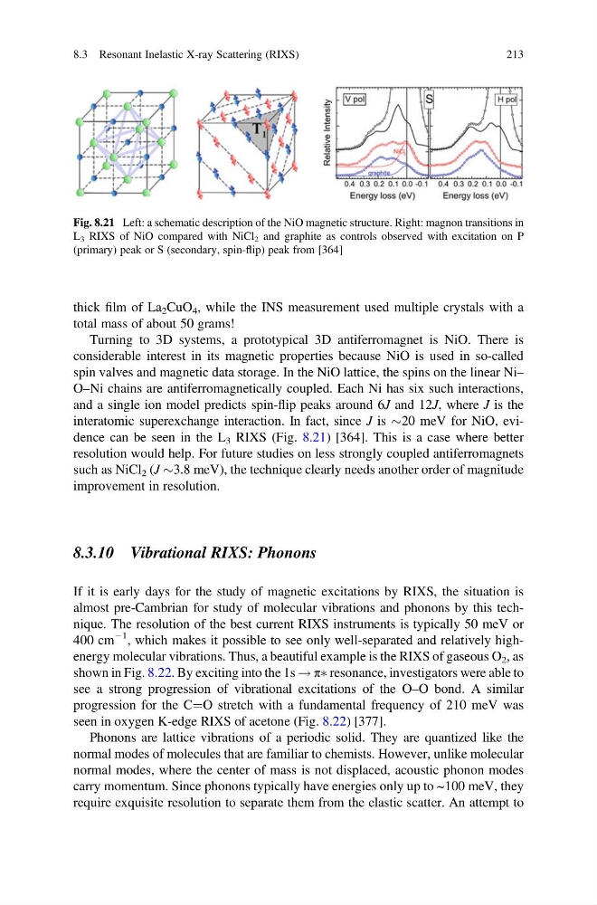
interatomic superexchange interaction. In fact, since J is $20 meV for NiO, evidence can be seen in the L 3 RIXS (Fig. 8.21) [364]. This is a case where better
resolution would help. For future studies on less strongly coupled antiferromagnets
such as NiCl 2 (J $3.8 meV), the technique clearly needs another order of magnitude
improvement in resolution.
8.3.10 Vibrational RIXS: Phonons
If it is early days for the study of magnetic excitations by RIXS, the situation is
almost pre-Cambrian for study of molecular vibrations and phonons by this technique. The resolution of the best current RIXS instruments is typically 50 meV or
400 cm
À1 , which makes it possible to see only well-separated and relatively highenergy molecular vibrations. Thus, a beautiful example is the RIXS of gaseous O 2 , as
shown in Fig. 8.22. By exciting into the 1s ! πà resonance, investigators were able to
see a strong progression of vibrational excitations of the O–O bond. A similar
progression for the C¼O stretch with a fundamental frequency of 210 meV was
seen in oxygen K-edge RIXS of acetone (Fig. 8.22) [377].
Phonons are lattice vibrations of a periodic solid. They are quantized like the
normal modes of molecules that are familiar to chemists. However, unlike molecular
normal modes, where the center of mass is not displaced, acoustic phonon modes
carry momentum. Since phonons typically have energies only up to ~100 meV, they
require exquisite resolution to separate them from the elastic scatter. An attempt to
Fig. 8.21 Left: a schematic description of the NiO magnetic structure. Right: magnon transitions in
L 3 RIXS of NiO compared with NiCl 2 and graphite as controls observed with excitation on P
(primary) peak or S (secondary, spin-flip) peak from [364]
8.3 Resonant Inelastic X-ray Scattering (RIXS)
213
total mass of about 50 grams!
Turning to 3D systems, a prototypical 3D antiferromagnet is NiO. There is
considerable interest in its magnetic properties because NiO is used in so-called
spin valves and magnetic data storage. In the NiO lattice, the spins on the linear Ni–
O–Ni chains are antiferromagnetically coupled. Each Ni has six such interactions,
and a single ion model predicts spin-flip peaks around 6J and 12J, where J is the
interatomic superexchange interaction. In fact, since J is $20 meV for NiO, evidence can be seen in the L 3 RIXS (Fig. 8.21) [364]. This is a case where better
resolution would help. For future studies on less strongly coupled antiferromagnets
such as NiCl 2 (J $3.8 meV), the technique clearly needs another order of magnitude
improvement in resolution.
8.3.10 Vibrational RIXS: Phonons
If it is early days for the study of magnetic excitations by RIXS, the situation is
almost pre-Cambrian for study of molecular vibrations and phonons by this technique. The resolution of the best current RIXS instruments is typically 50 meV or
400 cm
À1 , which makes it possible to see only well-separated and relatively highenergy molecular vibrations. Thus, a beautiful example is the RIXS of gaseous O 2 , as
shown in Fig. 8.22. By exciting into the 1s ! πà resonance, investigators were able to
see a strong progression of vibrational excitations of the O–O bond. A similar
progression for the C¼O stretch with a fundamental frequency of 210 meV was
seen in oxygen K-edge RIXS of acetone (Fig. 8.22) [377].
Phonons are lattice vibrations of a periodic solid. They are quantized like the
normal modes of molecules that are familiar to chemists. However, unlike molecular
normal modes, where the center of mass is not displaced, acoustic phonon modes
carry momentum. Since phonons typically have energies only up to ~100 meV, they
require exquisite resolution to separate them from the elastic scatter. An attempt to
Fig. 8.21 Left: a schematic description of the NiO magnetic structure. Right: magnon transitions in
L 3 RIXS of NiO compared with NiCl 2 and graphite as controls observed with excitation on P
(primary) peak or S (secondary, spin-flip) peak from [364]
8.3 Resonant Inelastic X-ray Scattering (RIXS)
213