

8.2.10 Applications
The chemical sensitivity of X-ray fluorescence leads to a variety of applications
beyond conventional elemental analysis. In some cases, one can simply examine the
fluorescence in detail for (a) chemical characterization. In another application, one
can (b) record an excitation spectrum while monitoring the fluorescence with “highenergy resolution fluorescence detection” in a technique abbreviated as “HERFD”
(see below) [344]. The HERFD approach can yield sharper excitation spectra at
absorption edges, and it can also allow EXAFS data to be recorded beyond interfering absorption edges, allowing so-called range-extended EXAFS [344]. Finally,
one can monitor different parts of the fluorescence while scanning the excitation
energy, yielding (c) site-selective and/or (d) spin-selective X-ray absorption data.
8.2.10.1 Chemical Characterization
We have already seen that different parts of the fluorescence spectrum are sensitive
to oxidation state, spin state, and the identity of neighboring ligands. Spin-state
diagnosis is a particularly nice application because the same sample can be compared
with itself at different temperatures or pressures. Rueff and coworkers have used the
1s3p Kβ X-ray emission spectral shape for the analysis of pressure-dependent
spectra of FeS [345], FeO [346], and Fe metal [347]. For FeS, the disappearance
of the Kβ´ satellite is a nice marker for the transition from high-spin to low-spin Fe
(Fig. 8.10). Similarly, the transition between the magnetic (bcc) and the nonmagnetic
(hcp) iron phases was seen from the shift of intensity toward the center of gravity.
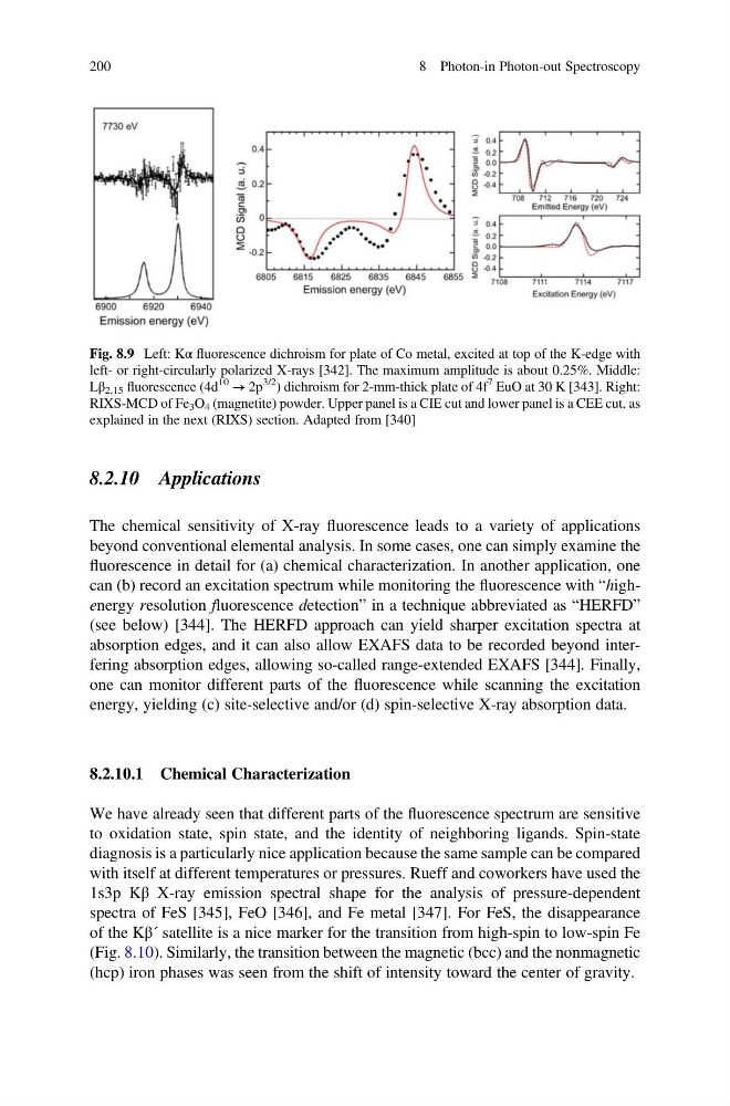
Fig. 8.9 Left: Kα fluorescence dichroism for plate of Co metal, excited at top of the K-edge with
left- or right-circularly polarized X-rays [342]. The maximum amplitude is about 0.25%. Middle:
Lβ 2,15 fluorescence (4d
10 → 2p
3/2
) dichroism for 2-mm-thick plate of 4f
7 EuO at 30 K [343]. Right:
RIXS-MCD of Fe 3 O 4 (magnetite) powder. Upper panel is a CIE cut and lower panel is a CEE cut, as
explained in the next (RIXS) section. Adapted from [340]
200
8 Photon-in Photon-out Spectroscopy
The chemical sensitivity of X-ray fluorescence leads to a variety of applications
beyond conventional elemental analysis. In some cases, one can simply examine the
fluorescence in detail for (a) chemical characterization. In another application, one
can (b) record an excitation spectrum while monitoring the fluorescence with “highenergy resolution fluorescence detection” in a technique abbreviated as “HERFD”
(see below) [344]. The HERFD approach can yield sharper excitation spectra at
absorption edges, and it can also allow EXAFS data to be recorded beyond interfering absorption edges, allowing so-called range-extended EXAFS [344]. Finally,
one can monitor different parts of the fluorescence while scanning the excitation
energy, yielding (c) site-selective and/or (d) spin-selective X-ray absorption data.
8.2.10.1 Chemical Characterization
We have already seen that different parts of the fluorescence spectrum are sensitive
to oxidation state, spin state, and the identity of neighboring ligands. Spin-state
diagnosis is a particularly nice application because the same sample can be compared
with itself at different temperatures or pressures. Rueff and coworkers have used the
1s3p Kβ X-ray emission spectral shape for the analysis of pressure-dependent
spectra of FeS [345], FeO [346], and Fe metal [347]. For FeS, the disappearance
of the Kβ´ satellite is a nice marker for the transition from high-spin to low-spin Fe
(Fig. 8.10). Similarly, the transition between the magnetic (bcc) and the nonmagnetic
(hcp) iron phases was seen from the shift of intensity toward the center of gravity.
Fig. 8.9 Left: Kα fluorescence dichroism for plate of Co metal, excited at top of the K-edge with
left- or right-circularly polarized X-rays [342]. The maximum amplitude is about 0.25%. Middle:
Lβ 2,15 fluorescence (4d
10 → 2p
3/2
) dichroism for 2-mm-thick plate of 4f
7 EuO at 30 K [343]. Right:
RIXS-MCD of Fe 3 O 4 (magnetite) powder. Upper panel is a CIE cut and lower panel is a CEE cut, as
explained in the next (RIXS) section. Adapted from [340]
200
8 Photon-in Photon-out Spectroscopy