

39
3
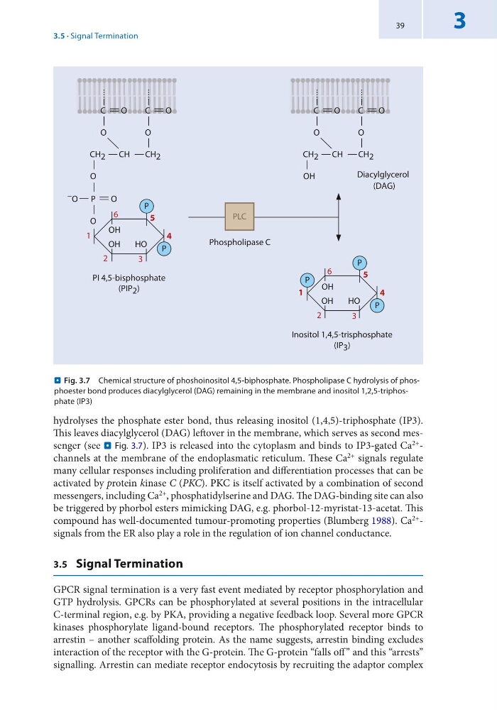
hydrolyses the phosphate ester bond, thus releasing inositol (1,4,5)-triphosphate (IP3).
This leaves diacylglycerol (DAG) leftover in the membrane, which serves as second messenger (see . Fig. 3.7). IP3 is released into the cytoplasm and binds to IP3-gated Ca 2+ -
channels at the membrane of the endoplasmatic reticulum. These Ca 2+ signals regulate
many cellular responses including proliferation and differentiation processes that can be
activated by protein kinase C (PKC). PKC is itself activated by a combination of second
messengers, including Ca 2+ , phosphatidylserine and DAG. The DAG-binding site can also
be triggered by phorbol esters mimicking DAG, e.g. phorbol-12-myristat-13-acetat. This
compound has well-documented tumour-promoting properties (Blumberg 1988). Ca 2+ -
signals from the ER also play a role in the regulation of ion channel conductance.
3.5 Signal Termination
GPCR signal termination is a very fast event mediated by receptor phosphorylation and
GTP hydrolysis. GPCRs can be phosphorylated at several positions in the intracellular
C-terminal region, e.g. by PKA, providing a negative feedback loop. Several more GPCR
kinases phosphorylate ligand-bound receptors. The phosphorylated receptor binds to
arrestin – another scaffolding protein. As the name suggests, arrestin binding excludes
interaction of the receptor with the G-protein. The G-protein “falls off ” and this “arrests”
signalling. Arrestin can mediate receptor endocytosis by recruiting the adaptor complex
Phospholipase C
C
O
O
CH 2
CH
CH 2
O
P
O
– O
O
OH
OH
P
4
5
PI 4,5-bisphosphate
(PIP 2 )
C
O
O
HO
P
C
O
O
CH 2
CH
CH 2
OH
C
O
O
OH
OH
P
4
5
Inositol 1,4,5-trisphosphate
(IP 3 )
HO
P
P
1
Diacylglycerol
(DAG)
2
3
6
1
2
3
6
PLC
. Fig. 3.7 Chemical structure of phoshoinositol 4,5-biphosphate. Phospholipase C hydrolysis of phosphoester bond produces diacylglycerol (DAG) remaining in the membrane and inositol 1,2,5-triphosphate (IP3)
3.5 · Signal Termination
3
hydrolyses the phosphate ester bond, thus releasing inositol (1,4,5)-triphosphate (IP3).
This leaves diacylglycerol (DAG) leftover in the membrane, which serves as second messenger (see . Fig. 3.7). IP3 is released into the cytoplasm and binds to IP3-gated Ca 2+ -
channels at the membrane of the endoplasmatic reticulum. These Ca 2+ signals regulate
many cellular responses including proliferation and differentiation processes that can be
activated by protein kinase C (PKC). PKC is itself activated by a combination of second
messengers, including Ca 2+ , phosphatidylserine and DAG. The DAG-binding site can also
be triggered by phorbol esters mimicking DAG, e.g. phorbol-12-myristat-13-acetat. This
compound has well-documented tumour-promoting properties (Blumberg 1988). Ca 2+ -
signals from the ER also play a role in the regulation of ion channel conductance.
3.5 Signal Termination
GPCR signal termination is a very fast event mediated by receptor phosphorylation and
GTP hydrolysis. GPCRs can be phosphorylated at several positions in the intracellular
C-terminal region, e.g. by PKA, providing a negative feedback loop. Several more GPCR
kinases phosphorylate ligand-bound receptors. The phosphorylated receptor binds to
arrestin – another scaffolding protein. As the name suggests, arrestin binding excludes
interaction of the receptor with the G-protein. The G-protein “falls off ” and this “arrests”
signalling. Arrestin can mediate receptor endocytosis by recruiting the adaptor complex
Phospholipase C
C
O
O
CH 2
CH
CH 2
O
P
O
– O
O
OH
OH
P
4
5
PI 4,5-bisphosphate
(PIP 2 )
C
O
O
HO
P
C
O
O
CH 2
CH
CH 2
OH
C
O
O
OH
OH
P
4
5
Inositol 1,4,5-trisphosphate
(IP 3 )
HO
P
P
1
Diacylglycerol
(DAG)
2
3
6
1
2
3
6
PLC
. Fig. 3.7 Chemical structure of phoshoinositol 4,5-biphosphate. Phospholipase C hydrolysis of phosphoester bond produces diacylglycerol (DAG) remaining in the membrane and inositol 1,2,5-triphosphate (IP3)
3.5 · Signal Termination