

2.4
Why Are Membrane Proteins Unstable in Detergent Solutions?
2.4.1
Instability of Detergent-Solubilized Membrane Proteins Is a General
Phenomenon
It is a sad fact of life, and the curse of the membrane biochemist, that most MPs start inactivating, more
or less rapidly, as soon as they are solubilized.
This is illustrated in Fig. 2.8, taking BR as an example (for an introduction to BR, see Chap. 1,
§ 1.6.1). When it is part of the purple membrane, its native environment, BR is extraordinarily stable
(see e.g. Brouillette et al. 1987): a suspension of purple membrane in water can be heated for 20 min at
70
C without any loss of the absorbance at ~554 nm that is characteristic of the native holoprotein
(Fig. 2.8A). On the contrary, once solubilized in n-octyl-β-D-thioglucoside (OTG; see Fig. 2.1), BR is
stable only until ~30
C, some signs of denaturation appearing already at 40
C: loss of absorbance at
554 nm and apparition of an absorbance peak, due to free retinal, around 380 nm, and of some
turbidity, due to the aggregation of the denatured protein (Fig. 2.8B). As expected, this increased
sensitivity to thermal denaturation translates into a shorter shelf life at constant temperature: whereas
purple membrane can be kept nearly indefinitely at room temperature, in the dark, as a suspension in
water, OTG-solubilized BR loses almost 30% of its absorbance at 554 nm after 1 day and is nearly
totally denatured after 6 days (Fig. 2.8C).
The case of BR is somewhat extreme given the extraordinary stability of this protein in its native
membrane, but, because of it, it is particularly telling. This behavior is very general. Very few MPs can
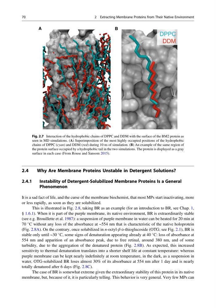
Fig. 2.7 Interaction of the hydrophobic chains of DPPC and DDM with the surface of the BM2 protein as
seen in MD simulations. (A) Superimposition of the most highly occupied positions of the hydrophobic
chains of DPPC (cyan) and DDM (red) during 10 ns of simulation. (B) An example of the same region of
the protein surface occupied by a hydrophobic tail in the two simulations. The protein is displayed as a gray
surface in each case (From Rouse and Sansom 2015).
70
2 Extracting Membrane Proteins from Their Native Environment
Why Are Membrane Proteins Unstable in Detergent Solutions?
2.4.1
Instability of Detergent-Solubilized Membrane Proteins Is a General
Phenomenon
It is a sad fact of life, and the curse of the membrane biochemist, that most MPs start inactivating, more
or less rapidly, as soon as they are solubilized.
This is illustrated in Fig. 2.8, taking BR as an example (for an introduction to BR, see Chap. 1,
§ 1.6.1). When it is part of the purple membrane, its native environment, BR is extraordinarily stable
(see e.g. Brouillette et al. 1987): a suspension of purple membrane in water can be heated for 20 min at
70
C without any loss of the absorbance at ~554 nm that is characteristic of the native holoprotein
(Fig. 2.8A). On the contrary, once solubilized in n-octyl-β-D-thioglucoside (OTG; see Fig. 2.1), BR is
stable only until ~30
C, some signs of denaturation appearing already at 40
C: loss of absorbance at
554 nm and apparition of an absorbance peak, due to free retinal, around 380 nm, and of some
turbidity, due to the aggregation of the denatured protein (Fig. 2.8B). As expected, this increased
sensitivity to thermal denaturation translates into a shorter shelf life at constant temperature: whereas
purple membrane can be kept nearly indefinitely at room temperature, in the dark, as a suspension in
water, OTG-solubilized BR loses almost 30% of its absorbance at 554 nm after 1 day and is nearly
totally denatured after 6 days (Fig. 2.8C).
The case of BR is somewhat extreme given the extraordinary stability of this protein in its native
membrane, but, because of it, it is particularly telling. This behavior is very general. Very few MPs can
Fig. 2.7 Interaction of the hydrophobic chains of DPPC and DDM with the surface of the BM2 protein as
seen in MD simulations. (A) Superimposition of the most highly occupied positions of the hydrophobic
chains of DPPC (cyan) and DDM (red) during 10 ns of simulation. (B) An example of the same region of
the protein surface occupied by a hydrophobic tail in the two simulations. The protein is displayed as a gray
surface in each case (From Rouse and Sansom 2015).
70
2 Extracting Membrane Proteins from Their Native Environment