

The crystals were then studied by neutron diffraction at different contrasts, so as to determine
the relative arrangements of the protein and detergent (Pebay-Peyroula et al. 1995). The contrast
match point of detergent polar heads (i.e. the D 2 O/H 2 O ratio at which their scattering length density is
equal to that of the solvent and their contribution to neutron scattering or diffraction vanishes; see
Chap. 9, Box 9.2) depends on their chemical composition and fraction of exchangeable hydrogens. In
the case of the polar head of OG, its contrast match point lies at ~52% D 2 O, so that at 40% D 2 O, its
contrast with the solution is weak, and it is mainly the octyl chains that contribute to building up the
diffraction pattern (see Timmins et al. 1994). Figure 2.5B shows the belt of OG surrounding the TM
region of OmpF in tetragonal crystals. This distribution is compared, in Fig. 2.5A, with that of
hydrophobic and aromatic residues (in black) at the surface of the protein (based on data in Cowan
et al. 1992). These residues form a belt ~2.5 nm high, which correspond to the expected thickness of
the acyl-chain region in Escherichia coli’s outer membrane. Comparison of the two sets of data
establishes that, as expected, the detergent substitutes for the lipids at the hydrophobic TM surface of
the protein, thus forming with it a water-soluble complex. The same conclusion has been reached in a
number of other neutron diffraction studies using other MPs, detergents, and/or crystal forms (see Roth
et al. 1989, 1991; Penel et al. 1998; Prince et al. 2003; Snijder et al. 2003), as well as in X-ray
diffraction studies in which the detergent belt was made visible by loading it under high pressure with
xenon or krypton (Sauer et al. 2002) or by increasing the electron density of the solvent (Norimatsu
et al. 2017).
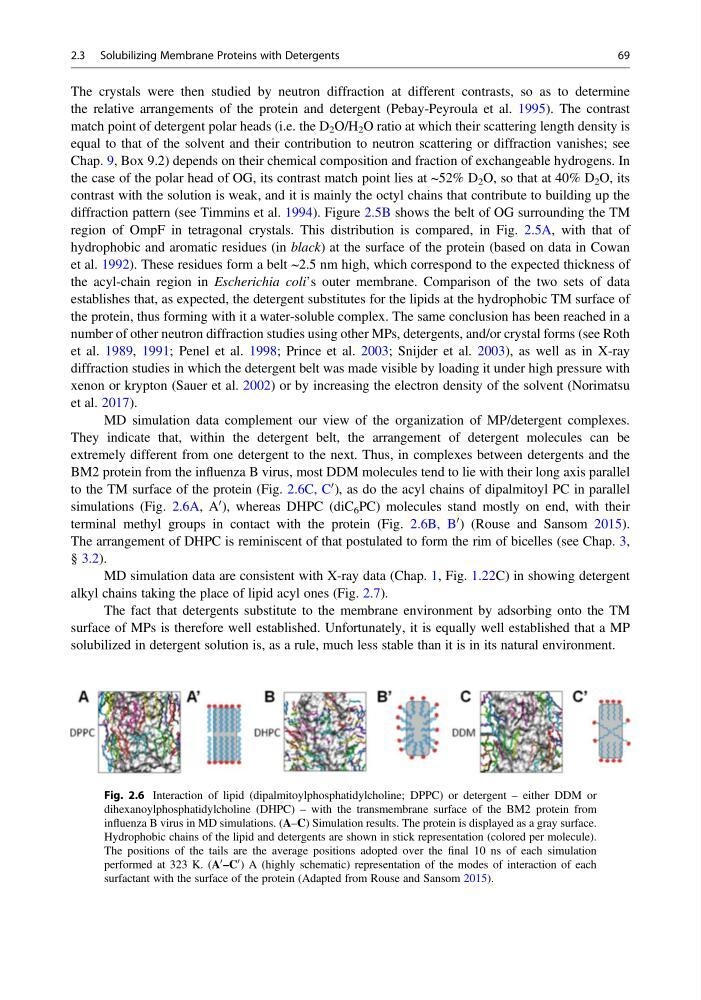
MD simulation data complement our view of the organization of MP/detergent complexes.
They indicate that, within the detergent belt, the arrangement of detergent molecules can be
extremely different from one detergent to the next. Thus, in complexes between detergents and the
BM2 protein from the influenza B virus, most DDM molecules tend to lie with their long axis parallel
to the TM surface of the protein (Fig. 2.6C, C
0 ), as do the acyl chains of dipalmitoyl PC in parallel
simulations (Fig. 2.6A, A
0 ), whereas DHPC (diC 6 PC) molecules stand mostly on end, with their
terminal methyl groups in contact with the protein (Fig. 2.6B, B
0 ) (Rouse and Sansom 2015).
The arrangement of DHPC is reminiscent of that postulated to form the rim of bicelles (see Chap. 3,
§ 3.2).
MD simulation data are consistent with X-ray data (Chap. 1, Fig. 1.22C) in showing detergent
alkyl chains taking the place of lipid acyl ones (Fig. 2.7).
The fact that detergents substitute to the membrane environment by adsorbing onto the TM
surface of MPs is therefore well established. Unfortunately, it is equally well established that a MP
solubilized in detergent solution is, as a rule, much less stable than it is in its natural environment.
Fig. 2.6 Interaction of lipid (dipalmitoylphosphatidylcholine; DPPC) or detergent – either DDM or
dihexanoylphosphatidylcholine (DHPC) – with the transmembrane surface of the BM2 protein from
influenza B virus in MD simulations. (A–C) Simulation results. The protein is displayed as a gray surface.
Hydrophobic chains of the lipid and detergents are shown in stick representation (colored per molecule).
The positions of the tails are the average positions adopted over the final 10 ns of each simulation
performed at 323 K. (A
0 –C
0 ) A (highly schematic) representation of the modes of interaction of each
surfactant with the surface of the protein (Adapted from Rouse and Sansom 2015).
2.3 Solubilizing Membrane Proteins with Detergents
69
the relative arrangements of the protein and detergent (Pebay-Peyroula et al. 1995). The contrast
match point of detergent polar heads (i.e. the D 2 O/H 2 O ratio at which their scattering length density is
equal to that of the solvent and their contribution to neutron scattering or diffraction vanishes; see
Chap. 9, Box 9.2) depends on their chemical composition and fraction of exchangeable hydrogens. In
the case of the polar head of OG, its contrast match point lies at ~52% D 2 O, so that at 40% D 2 O, its
contrast with the solution is weak, and it is mainly the octyl chains that contribute to building up the
diffraction pattern (see Timmins et al. 1994). Figure 2.5B shows the belt of OG surrounding the TM
region of OmpF in tetragonal crystals. This distribution is compared, in Fig. 2.5A, with that of
hydrophobic and aromatic residues (in black) at the surface of the protein (based on data in Cowan
et al. 1992). These residues form a belt ~2.5 nm high, which correspond to the expected thickness of
the acyl-chain region in Escherichia coli’s outer membrane. Comparison of the two sets of data
establishes that, as expected, the detergent substitutes for the lipids at the hydrophobic TM surface of
the protein, thus forming with it a water-soluble complex. The same conclusion has been reached in a
number of other neutron diffraction studies using other MPs, detergents, and/or crystal forms (see Roth
et al. 1989, 1991; Penel et al. 1998; Prince et al. 2003; Snijder et al. 2003), as well as in X-ray
diffraction studies in which the detergent belt was made visible by loading it under high pressure with
xenon or krypton (Sauer et al. 2002) or by increasing the electron density of the solvent (Norimatsu
et al. 2017).
MD simulation data complement our view of the organization of MP/detergent complexes.
They indicate that, within the detergent belt, the arrangement of detergent molecules can be
extremely different from one detergent to the next. Thus, in complexes between detergents and the
BM2 protein from the influenza B virus, most DDM molecules tend to lie with their long axis parallel
to the TM surface of the protein (Fig. 2.6C, C
0 ), as do the acyl chains of dipalmitoyl PC in parallel
simulations (Fig. 2.6A, A
0 ), whereas DHPC (diC 6 PC) molecules stand mostly on end, with their
terminal methyl groups in contact with the protein (Fig. 2.6B, B
0 ) (Rouse and Sansom 2015).
The arrangement of DHPC is reminiscent of that postulated to form the rim of bicelles (see Chap. 3,
§ 3.2).
MD simulation data are consistent with X-ray data (Chap. 1, Fig. 1.22C) in showing detergent
alkyl chains taking the place of lipid acyl ones (Fig. 2.7).
The fact that detergents substitute to the membrane environment by adsorbing onto the TM
surface of MPs is therefore well established. Unfortunately, it is equally well established that a MP
solubilized in detergent solution is, as a rule, much less stable than it is in its natural environment.
Fig. 2.6 Interaction of lipid (dipalmitoylphosphatidylcholine; DPPC) or detergent – either DDM or
dihexanoylphosphatidylcholine (DHPC) – with the transmembrane surface of the BM2 protein from
influenza B virus in MD simulations. (A–C) Simulation results. The protein is displayed as a gray surface.
Hydrophobic chains of the lipid and detergents are shown in stick representation (colored per molecule).
The positions of the tails are the average positions adopted over the final 10 ns of each simulation
performed at 323 K. (A
0 –C
0 ) A (highly schematic) representation of the modes of interaction of each
surfactant with the surface of the protein (Adapted from Rouse and Sansom 2015).
2.3 Solubilizing Membrane Proteins with Detergents
69