

peptide once each partner has the option of integrating into a membrane, where lipids compete with
APols for the hydrophobic surface of the peptide and entropy works to separate the two partners.
Förster resonance energy transfer experiments would permit direct probing of this issue. They have not
been carried out in Study 15.1. However, as discussed in more detail in Chap. 5, § 5.7, and 11, § 11.3.2,
indirect evidence in favor of such a dissociation is provided by the experiments of Polovinkin et al.
(2014), in which bacteriorhodopsin/A8-35 complexes were mixed with a lipidic mesophase. The
protein diffused and crystallized in the mesophase, without being accompanied by any APol (see
Chap. 11, § 11.3.2). It is most likely that, in the experiment shown in Fig. 15.7, the two partners
separated upon binding to the plasma membrane and diffused separately at the surface of the cell
before being endocytosed. Whether internalized separately or not, they ended up in the same endocytic
compartments, as shown by the images collected after 72 h.
15.4 Biodistribution and Elimination of Amphipols Following Injection
in Mice
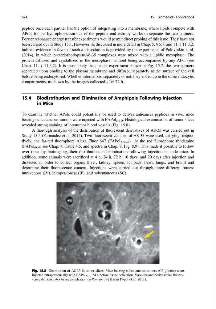
To examine whether APols could potentially be used to deliver anticancer peptides in vivo, mice
bearing subcutaneous tumors were injected with FAPol NBD . Histological examination of tumor slices
revealed strong staining of intratumor blood vessels (Fig. 15.8).
A thorough analysis of the distribution of fluorescent derivatives of A8-35 was carried out in
Study 15.5 (Fernandez et al. 2014). Two fluorescent versions of A8-35 were used, carrying, respectively, the far-red fluorophore Alexa Fluor 647 (FAPol AF647 ) or the red fluorophore rhodamine
(FAPol rhod ; see Chap. 4, Table 4.5, and spectra in Chap. 8, Fig. 8.9). This made it possible to follow
over time, by bioimaging, their distribution and elimination following injection in nude mice. In
addition, some animals were sacrificed at 4 h, 24 h, 72 h, 10 days, and 20 days after injection and
dissected in order to collect organs (liver, kidney, spleen, fat pads, heart, lungs, and brain) and
determine their fluorescence content. Injections were carried out through three different routes:
intravenous (IV), intraperitoneal (IP), and subcutaneous (SC).
Fig. 15.8 Distribution of A8-35 in tumor slices. Mice bearing subcutaneous tumors (C6 glioma) were
injected intraperitoneally with FAPol NBD 24 h before tissue collection. Vascular and perivascular fluorescence demonstrates tissue penetration (yellow arrow) (From Popot et al. 2011).
674
15 Biomedical Applications