

network supporting tumor growth by hybridizing it with TM peptides largely reduces brain and breast
tumor expansion and dissemination both in vitro and in vivo (Nasarre et al. 2010; Arpel et al. 2016).
Similar results have been achieved against glioblastoma using a TM peptide derived from Plexin-A1
(Jacob et al. 2016). TM peptides, however, are insoluble and prone to aggregation, which makes it
difficult to ensure optimal distribution and efficacy in whole organisms. As described in Chap. 5
(Table 5.1), APols have been shown to keep various TM peptides soluble (Gohon 1996; Duarte et al.
2008; Stangl et al. 2014), suggesting that they could be used as solubilizing and vectorizing agents.
This has led to examining the ability of APols to distribute throughout the organism of mice and to
deliver TM peptides to cells in culture.
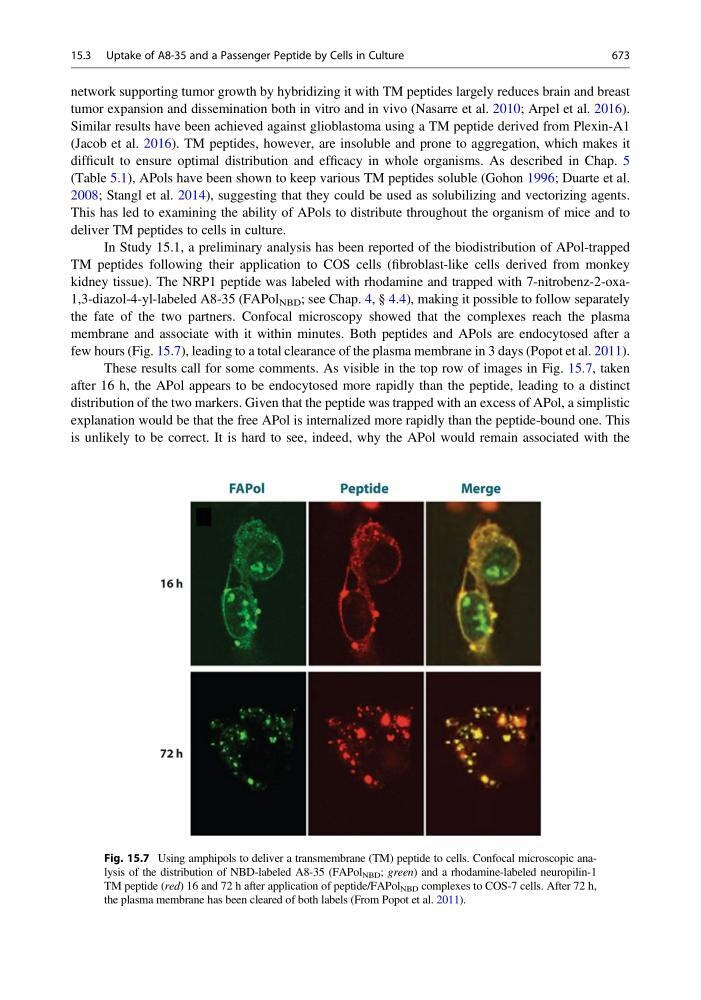
In Study 15.1, a preliminary analysis has been reported of the biodistribution of APol-trapped
TM peptides following their application to COS cells (fibroblast-like cells derived from monkey
kidney tissue). The NRP1 peptide was labeled with rhodamine and trapped with 7-nitrobenz-2-oxa1,3-diazol-4-yl-labeled A8-35 (FAPol NBD ; see Chap. 4, § 4.4), making it possible to follow separately
the fate of the two partners. Confocal microscopy showed that the complexes reach the plasma
membrane and associate with it within minutes. Both peptides and APols are endocytosed after a
few hours (Fig. 15.7), leading to a total clearance of the plasma membrane in 3 days (Popot et al. 2011).
These results call for some comments. As visible in the top row of images in Fig. 15.7, taken
after 16 h, the APol appears to be endocytosed more rapidly than the peptide, leading to a distinct
distribution of the two markers. Given that the peptide was trapped with an excess of APol, a simplistic
explanation would be that the free APol is internalized more rapidly than the peptide-bound one. This
is unlikely to be correct. It is hard to see, indeed, why the APol would remain associated with the
Fig. 15.7 Using amphipols to deliver a transmembrane (TM) peptide to cells. Confocal microscopic analysis of the distribution of NBD-labeled A8-35 (FAPol NBD ; green) and a rhodamine-labeled neuropilin-1
TM peptide (red) 16 and 72 h after application of peptide/FAPol NBD complexes to COS-7 cells. After 72 h,
the plasma membrane has been cleared of both labels (From Popot et al. 2011).
15.3 Uptake of A8-35 and a Passenger Peptide by Cells in Culture
673