

The nature of the problems encountered when attempting to identify all proteins from a tissue or
cell culture, or a membrane fraction thereof, is summarized in Ning et al. (2013). In brief, the study of
the proteome by mass spectrometry requires the lysis and recovery of proteins from cells and tissues.
This requires the disruption of plasma and intracellular membranes, in order to release intracellular and
intraorganelle proteins. In addition, most proteins associated with membranes, be it through TM
domains, non-covalent interactions with TM proteins or lipids, or anchoring side groups (see
Chap. 1), are insoluble in water. Detergents are generally used to dissolve membranes and keep
hydrophobic proteins water-soluble. Detergents, however, at the concentration used to disrupt
membranes, tend to interfere with the proteolytic enzymes, such as trypsin, used to produce peptides
for MS analysis. They must therefore be removed prior to proteolysis. Furthermore, most of them
interfere with the chromatography and MS of peptides by high-pressure LC/ESI tandem mass
spectrometry (HPLC-ESI-MS). Removing the detergent causes most MPs to precipitate. Chaotrophic
agents are therefore required to keep them soluble for the reduction, alkylation, and proteolysis steps.
The activity of proteolytic enzymes is typically tolerant of low concentration of chaotropes. However,
most chaotropic agents are not suitable for MS and, therefore, have to be removed following
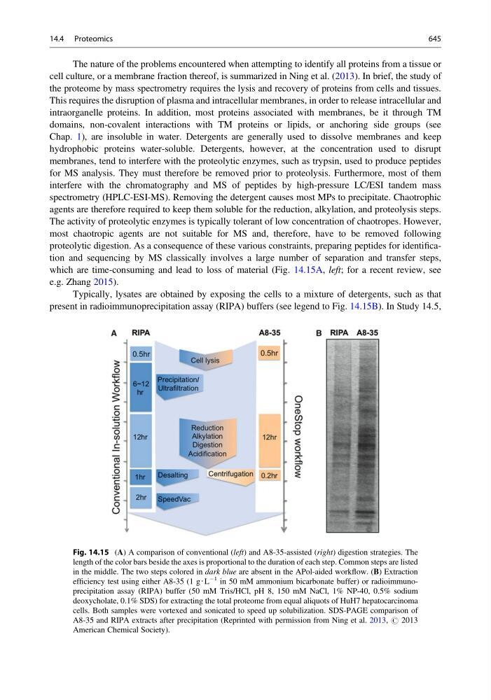
proteolytic digestion. As a consequence of these various constraints, preparing peptides for identification and sequencing by MS classically involves a large number of separation and transfer steps,
which are time-consuming and lead to loss of material (Fig. 14.15A, left; for a recent review, see
e.g. Zhang 2015).
Typically, lysates are obtained by exposing the cells to a mixture of detergents, such as that
present in radioimmunoprecipitation assay (RIPA) buffers (see legend to Fig. 14.15B). In Study 14.5,
Fig. 14.15 (A) A comparison of conventional (left) and A8-35-assisted (right) digestion strategies. The
length of the color bars beside the axes is proportional to the duration of each step. Common steps are listed
in the middle. The two steps colored in dark blue are absent in the APol-aided workflow. (B) Extraction
efficiency test using either A8-35 (1 g‧L
À1 in 50 mM ammonium bicarbonate buffer) or radioimmunoprecipitation assay (RIPA) buffer (50 mM Tris/HCl, pH 8, 150 mM NaCl, 1% NP-40, 0.5% sodium
deoxycholate, 0.1% SDS) for extracting the total proteome from equal aliquots of HuH7 hepatocarcinoma
cells. Both samples were vortexed and sonicated to speed up solubilization. SDS-PAGE comparison of
A8-35 and RIPA extracts after precipitation (Reprinted with permission from Ning et al. 2013, # 2013
American Chemical Society).
14.4 Proteomics
645