

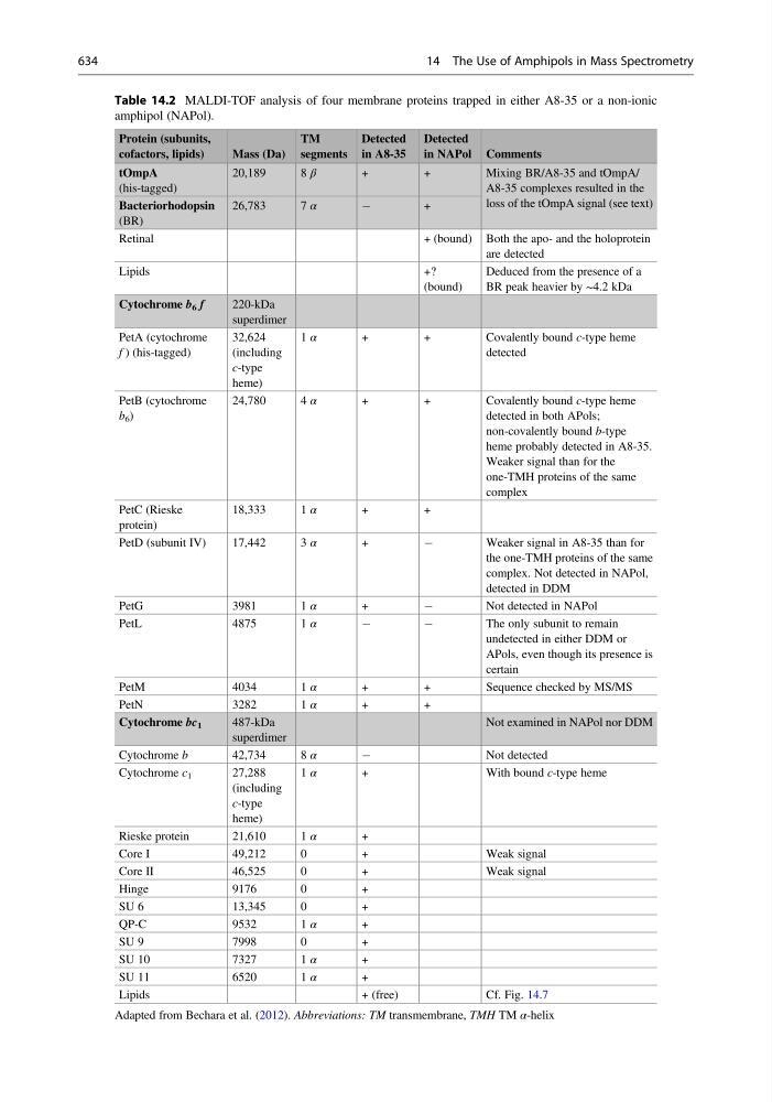
Table 14.2 MALDI-TOF analysis of four membrane proteins trapped in either A8-35 or a non-ionic
amphipol (NAPol).
Protein (subunits,
cofactors, lipids)
Mass (Da)
TM
segments
Detected
in A8-35
Detected
in NAPol Comments
tOmpA
(his-tagged)
20,189
8 β
+
+
Mixing BR/A8-35 and tOmpA/
A8-35 complexes resulted in the
loss of the tOmpA signal (see text)
Bacteriorhodopsin
(BR)
26,783
7 α
À
+
Retinal
+ (bound) Both the apo- and the holoprotein
are detected
Lipids
+?
(bound)
Deduced from the presence of a
BR peak heavier by ~4.2 kDa
Cytochrome b 6 f
220-kDa
superdimer
PetA (cytochrome
f ) (his-tagged)
32,624
(including
c-type
heme)
1 α
+
+
Covalently bound c-type heme
detected
PetB (cytochrome
b 6 )
24,780
4 α
+
+
Covalently bound c-type heme
detected in both APols;
non-covalently bound b-type
heme probably detected in A8-35.
Weaker signal than for the
one-TMH proteins of the same
complex
PetC (Rieske
protein)
18,333
1 α
+
+
PetD (subunit IV)
17,442
3 α
+
À
Weaker signal in A8-35 than for
the one-TMH proteins of the same
complex. Not detected in NAPol,
detected in DDM
PetG
3981
1 α
+
À
Not detected in NAPol
PetL
4875
1 α
À
À
The only subunit to remain
undetected in either DDM or
APols, even though its presence is
certain
PetM
4034
1 α
+
+
Sequence checked by MS/MS
PetN
3282
1 α
+
+
Cytochrome bc 1
487-kDa
superdimer
Not examined in NAPol nor DDM
Cytochrome b
42,734
8 α
À
Not detected
Cytochrome c 1
27,288
(including
c-type
heme)
1 α
+
With bound c-type heme
Rieske protein
21,610
1 α
+
Core I
49,212
0
+
Weak signal
Core II
46,525
0
+
Weak signal
Hinge
9176
0
+
SU 6
13,345
0
+
QP-C
9532
1 α
+
SU 9
7998
0
+
SU 10
7327
1 α
+
SU 11
6520
1 α
+
Lipids
+ (free)
Cf. Fig. 14.7
Adapted from Bechara et al. (2012). Abbreviations: TM transmembrane, TMH TM α-helix
634
14 The Use of Amphipols in Mass Spectrometry