

naked protein, but also to a collection of heavier ions that are too light to contain the APol and most
likely correspond to BR/lipid complexes. The rate of proteolysis decreased in the order DDM ! A8-35
> NAPols, which correlates with the stabilizing potential of the three types of surfactants (see Chap. 5,
§ 5.5). These observations suggest that resorting to APols does not improve the sequence coverage of
MPs, but may possibly help in distinguishing between cleavage sites that are always exposed and sites
that become so only upon conformational excursions of the protein.
Most of the subunits present in cytochrome b 6 f were detected whatever the APol used. However,
subunits PetD (three TM α-helices) and PetG (one TM helix) were not detected in the NAPol-trapped
preparation. Subunit PetL, although known to be present (Stroebel et al. 2003), was detected neither in
A8-35, NAPol, nor DDM. Limited tryptic digestion did not show any significant surfactant-dependent
differences between samples in terms of the fragments obtained. In terms of kinetics, however, peptide
digests appeared more quickly when the b 6 f complex was solubilized in DDM as compared to being
trapped in A8-35. As observed with BR, the b 6 f was most resistant to tryptic digestion when trapped in
NAPols, only a few peptides being obtained from such samples. All tryptic peptides that were
identified by MS/MS belonged to the three largest subunits, namely cytochromes f and b 6 and, to a
lesser extent, the Rieske protein. This is not surprising, given that those are the subunits most exposed
to the aqueous solution. Subunit IV is mostly buried, while the small subunits PetG, PetL, PetM, and
PetN are short, mainly transmembrane peptides, with no or few arginyl or lysyl residues (see Stroebel
et al. 2003). Intact small subunits were occasionally detected in digested samples that had not been
micropurified.
Cytochrome bc 1 was analyzed only in A8-35. Ten out of the 11 subunits were detected. The
undetected subunit is cytochrome b, which comprises eight TM helices (see Berry et al. 2000). This is
reminiscent of the fact that, in the case of the b 6 f complex, the two subunits with multiple TM helices,
cytochrome b 6 and subunit IV, yield weaker signals than those with a single TM helix. As for the
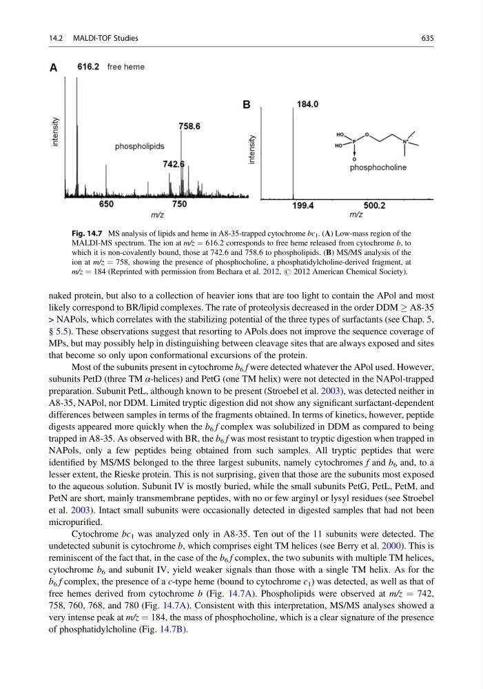
b 6 f complex, the presence of a c-type heme (bound to cytochrome c 1 ) was detected, as well as that of
free hemes derived from cytochrome b (Fig. 14.7A). Phospholipids were observed at m/z ¼ 742,
758, 760, 768, and 780 (Fig. 14.7A). Consistent with this interpretation, MS/MS analyses showed a
very intense peak at m/z ¼ 184, the mass of phosphocholine, which is a clear signature of the presence
of phosphatidylcholine (Fig. 14.7B).
Fig. 14.7 MS analysis of lipids and heme in A8-35-trapped cytochrome bc 1 . (A) Low-mass region of the
MALDI-MS spectrum. The ion at m/z ¼ 616.2 corresponds to free heme released from cytochrome b, to
which it is non-covalently bound, those at 742.6 and 758.6 to phospholipids. (B) MS/MS analysis of the
ion at m/z ¼ 758, showing the presence of phosphocholine, a phosphatidylcholine-derived fragment, at
m/z ¼ 184 (Reprinted with permission from Bechara et al. 2012, # 2012 American Chemical Society).
14.2 MALDI-TOF Studies
635