

ankyrin repeat proteins” (DARPins) (Binz et al. 2003; Stumpp et al. 2003; Sennhauser and Grütter
2008). Other types of repeats based on Armadillo, leucine-rich repeats, or tetratricopeptide repeats
have also produced specific binders (Boersma and Plückthun 2011).
Philippe Minard and coworkers have introduced a new family of artificial repeat proteins named
“αReps.” αReps are derived from a subgroup of HEAT-like repeats found in several cytoplasmic
proteins, the names of four of which are at the origin of the acronym (Huntingtin, elongation factor
3 (EF3), protein phosphatase 2A (PP2A), and the yeast kinase TOR1). αReps are efficiently expressed
and folded, and they are very stable. Large libraries have been generated (Guellouz et al. 2013), from
which tight and specific binders have been selected against several soluble proteins, with dissociation
constants, K D , in the nanomolar to micromolar range. Crystallographic structures of αRep/target
protein complexes show conformation-specific recognition (Guellouz et al. 2013).
For the selection of binding proteins as putative crystallization helpers, it is critical that, at all
stages of the selection process, the purified MPs used as targets be immobilized on a solid support
under their native state. In Studies 13.3/13.6, biotinylated APols were used to this end. The approach
was tested with four model MPs of known structures, MexB and OprM from the Gram
À bacterium
Pseudomonas aeruginosa efflux pump system, the cytochrome bc 1 complex from beef heart
mitochondria, and BR from H. salinarum. MexB and OprM had not been previously trapped with
APols, whereas BR and the bc 1 complex had already been used in APol-mediated immobilization
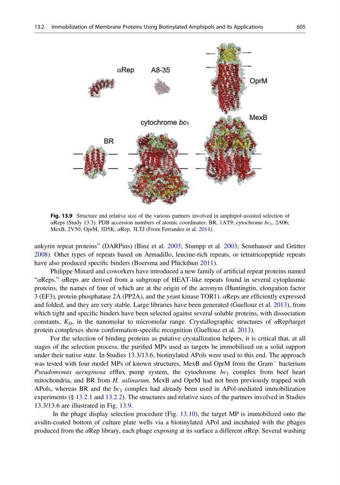
experiments (§ 13.2.1 and 13.2.2). The structures and relative sizes of the partners involved in Studies
13.3/13.6 are illustrated in Fig. 13.9.
In the phage display selection procedure (Fig. 13.10), the target MP is immobilized onto the
avidin-coated bottom of culture plate wells via a biotinylated APol and incubated with the phages
produced from the αRep library, each phage exposing at its surface a different αRep. Several washing
Fig. 13.9 Structure and relative size of the various partners involved in amphipol-assisted selection of
αReps (Study 13.3). PDB accession numbers of atomic coordinates: BR, 1AT9; cytochrome bc 1 , 2A06;
MexB, 2V50; OprM, 3D5K; αRep, 3LTJ (From Ferrandez et al. 2014).
13.2 Immobilization of Membrane Proteins Using Biotinylated Amphipols and Its Applications
605