

unfunctionalized A8-35 and FAPol AF647 also showed a signal on the avidin-decorated surface,
whereas no fluorescence signal was detected on the electrodes bearing SA or neutravidin (Della Pia
et al. 2014a). The higher non-specific binding to the avidin surface can likely be attributed to
electrostatic interactions taking place in the low ionic strength buffer solution. A8-35-trapped BR
bears a high negative charge due to the many carboxylates carried by the APol belt, all of which are
ionized at pH 7.2 (Gohon et al. 2004; see Chap. 4, § 4.3.1.2.2). As a result, it adsorbs non-specifically
onto the positively charged avidin substrate (isoelectric point $10.5). In contrast, SA- and neutravidinmodified surfaces have a slightly negative charge at pH 7.2 (isoelectric points 6.1 and 6.3, respectively;
Petrlova et al. 2007) and exhibit much less non-specific binding of BR/A8-35 complexes than avidin.
Non-specific binding of cationic ligands by BA8-35 is a recurrent concern (Ferrandez et al. 2014; Mary
et al. 2014, and unpublished data by various groups), which has led to the development of biotinylated
non-ionic APols, as will be described in § 13.2.3.
13.2.3 Using Ionic vs. Non-ionic Biotinylated Amphipols for Phage Display
Selection of Membrane Protein Binders
Resorting to specific and tight-binding protein partners can considerably facilitate the challenging
process of MP crystallization (cf. Chap. 11, § 11.2.2). The binders are sometimes referred to as
“crystallization chaperones” (Koide 2009; Lieberman et al. 2011; not necessarily a perfectly appropriate term given that they are part of the final crystals, and not temporary facilitators as chaperones are
supposed to be), or “orthoses” (Ferrandez et al. 2014). Natural binders include monoclonal antibody F v
fragments, an approach pioneered by Hartmut Michel (Ostermeier et al. 1995), heterologously
produced versions thereof, and camelid nanobodies. Among the many types of artificial binders that
have been proposed (for a general review, see Skerra 2007), a very promising class based on repeat
proteins has been introduced by Andreas Plückthun and colleagues under the name of “designed
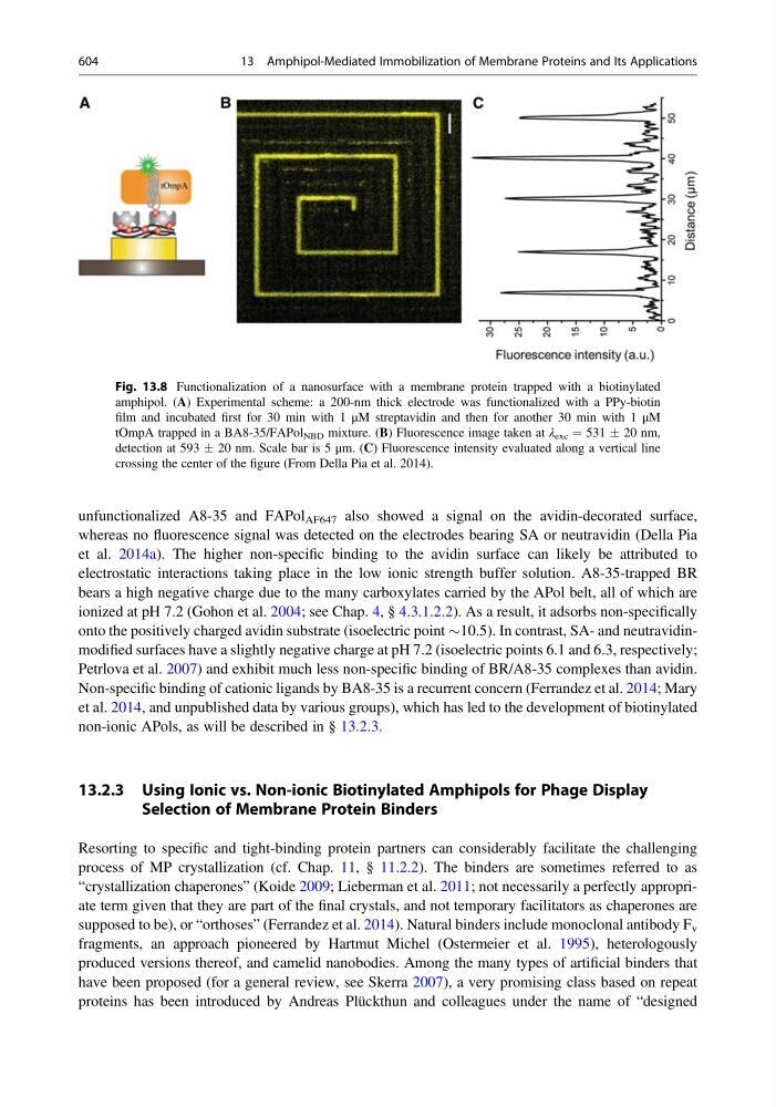
Fig. 13.8 Functionalization of a nanosurface with a membrane protein trapped with a biotinylated
amphipol. (A) Experimental scheme: a 200-nm thick electrode was functionalized with a PPy-biotin
film and incubated first for 30 min with 1 μM streptavidin and then for another 30 min with 1 μM
tOmpA trapped in a BA8-35/FAPol NBD mixture. (B) Fluorescence image taken at λ exc ¼ 531 Æ 20 nm,
detection at 593 Æ 20 nm. Scale bar is 5 μm. (C) Fluorescence intensity evaluated along a vertical line
crossing the center of the figure (From Della Pia et al. 2014).
604
13 Amphipol-Mediated Immobilization of Membrane Proteins and Its Applications