

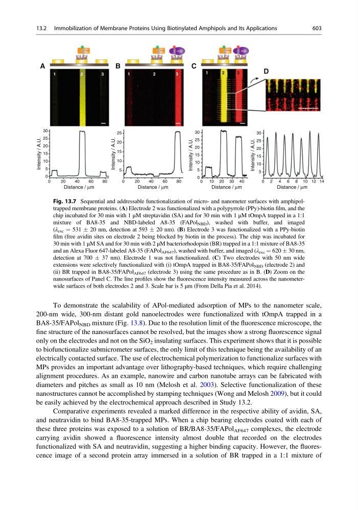
To demonstrate the scalability of APol-mediated adsorption of MPs to the nanometer scale,
200-nm wide, 300-nm distant gold nanoelectrodes were functionalized with tOmpA trapped in a
BA8-35/FAPol NBD mixture (Fig. 13.8). Due to the resolution limit of the fluorescence microscope, the
fine structure of the nanosurfaces cannot be resolved, but the images show a strong fluorescence signal
only on the electrodes and not on the SiO 2 insulating surfaces. This experiment shows that it is possible
to biofunctionalize submicrometer surfaces, the only limit of this technique being the availability of an
electrically contacted surface. The use of electrochemical polymerization to functionalize surfaces with
MPs provides an important advantage over lithography-based techniques, which require challenging
alignment procedures. As an example, nanowire and carbon nanotube arrays can be fabricated with
diameters and pitches as small as 10 nm (Melosh et al. 2003). Selective functionalization of these
nanostructures cannot be accomplished by stamping techniques (Wong and Melosh 2009), but it could
be easily achieved by the electrochemical approach described in Study 13.2.
Comparative experiments revealed a marked difference in the respective ability of avidin, SA,
and neutravidin to bind BA8-35-trapped MPs. When a chip bearing electrodes coated with each of
these three proteins was exposed to a solution of BR/BA8-35/FAPol AF647 complexes, the electrode
carrying avidin showed a fluorescence intensity almost double that recorded on the electrodes
functionalized with SA and neutravidin, suggesting a higher binding capacity. However, the fluorescence image of a second protein array immersed in a solution of BR trapped in a 1:1 mixture of
0
5
10
15
20
25
5
10
15
20
25
1
2
3
1
1
2
3
2
3
30
A
B
C
D
0
2 0
4 0
Distance / µm
Intensity / A.U.
0
5
10
15
20
25
30
Intensity / A.U.
5
10
15
20
25
30
Intensity / A.U.
Intensity / A.U.
60
80
0
20
40
Distance / µm
60
80
0
0 2 4 6 8 10 12 14
10
40
30
20
Distance / µm
Distance / µm
Fig. 13.7 Sequential and addressable functionalization of micro- and nanometer surfaces with amphipoltrapped membrane proteins. (A) Electrode 2 was functionalized with a polypyrrole (PPy)-biotin film, and the
chip incubated for 30 min with 1 μM streptavidin (SA) and for 30 min with 1 μM tOmpA trapped in a 1:1
mixture of BA8-35 and NBD-labeled A8-35 (FAPol NBD ), washed with buffer, and imaged
(λ exc ¼ 531 Æ 20 nm, detection at 593 Æ 20 nm). (B) Electrode 3 was functionalized with a PPy-biotin
film (free avidin sites on electrode 2 being blocked by biotin in the process). The chip was incubated for
30 min with 1 μM SA and for 30 min with 2 μM bacteriorhodopsin (BR) trapped in a 1:1 mixture of BA8-35
and an Alexa Fluor 647-labeled A8-35 (FAPol AF647 ), washed with buffer, and imaged (λ exc ¼ 620 Æ 30 nm,
detection at 700 Æ 37 nm). Electrode 1 was not functionalized. (C) Two electrodes with 50 nm wide
extensions were selectively functionalized with (i) tOmpA trapped in BA8-35/FAPol NBD (electrode 2) and
(ii) BR trapped in BA8-35/FAPol AF647 (electrode 3) using the same procedure as in B. (D) Zoom on the
nanosurfaces of Panel C. The line profiles show the fluorescence intensity measured across the nanometerwide surfaces of both electrodes 2 and 3. Scale bar is 5 μm (From Della Pia et al. 2014).
13.2 Immobilization of Membrane Proteins Using Biotinylated Amphipols and Its Applications
603