

on electrode 3 (in which process the remaining free avidin sites on the chip were blocked by biotin) and
the chip exposed successively first to SA and then to BR/BA8-35/FAPol A647 complexes. As shown in
Fig. 13.7B, the procedure resulted in MP adsorption onto electrode 3 only, with minimal contamination of the electrode that had not been functionalized (electrode 1), or of that where a MP had already
been immobilized and the biotin-binding sites blocked (electrode 2). These results further demonstrate
that BA8-35-trapped MPs remain attached after incubation with the monomeric solution of pyrrole and
are not detached by the application of the electric field needed for the activation of the adjacent
electrode, nor of the second solution of MP/APol complexes. The procedure ought to be scalable to any
number of different MPs and/or other biotin-tagged compounds.
Because of the highly localized nature of electric fields, this approach can be implemented on
submicrometer electrodes. This is shown in Fig. 13.7C, D, where two electrodes with features as small
as $50 nm were functionalized with BR and tOmpA.
A
B
1
2
3
1
2
3
1
2
3
C
0
0
10 20 30 40
Distance / µm
Intensity / A.U.
50 60 70
2
4
6
8
10
0
0
10 20 30 40
Distance / µm
Intensity / A.U.
50 60 70
2
4
6
8
10
4
6
8
10
12
14
16
18
+
0
10 20 30 40
Distance / µm
Intensity / A.U.
50 60 70
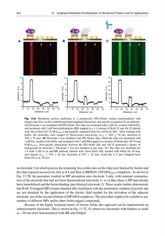
Fig. 13.6 Membrane protein anchoring to a polypyrrole (PPy)-biotin surface functionalized with
streptavidin (SA) via the avidin/biotinylated amphipol interaction, and specific recognition by an antibody.
(A) Electrode 1 was modified with PPy-biotin. The chip was incubated with 1 μM SA, washed with buffer,
and incubated with 1 μM bacteriorhodopsin (BR) trapped in a 1:1 mixture of BA8-35 and A8-35 labeled
with Alexa Fluor 647 (FAPol AF647 ) and partially separated from free APols by SEC. After washing with
buffer, the electrodes were imaged by fluorescence microscopy (λ exc ¼ 620 Æ 30 nm, detection at
700 Æ 35 nm). (B) Electrode 2 was modified with PPy-biotin, after which the chip was incubated with
1 μM SA, washed with buffer, and incubated with 1 μM BR trapped in a mixture of biotin-free A8-35 and
FAPol AF647 . Non-specific interactions between the PPy-biotin film and A8-35 generated a degree of
background on electrode 2. Electrode 3 was not modified at any time. (C) The chip was incubated for
1 h with 5 nM of an anti-BR antibody labeled with Alexa Fluor 488, washed with buffer for 30 min,
and imaged (λ exc ¼ 470 Æ 20 nm, detection at 525 Æ 25 nm). Scale bar is 5 μm (Adapted from
Della Pia et al. 2014a).
602
13 Amphipol-Mediated Immobilization of Membrane Proteins and Its Applications