

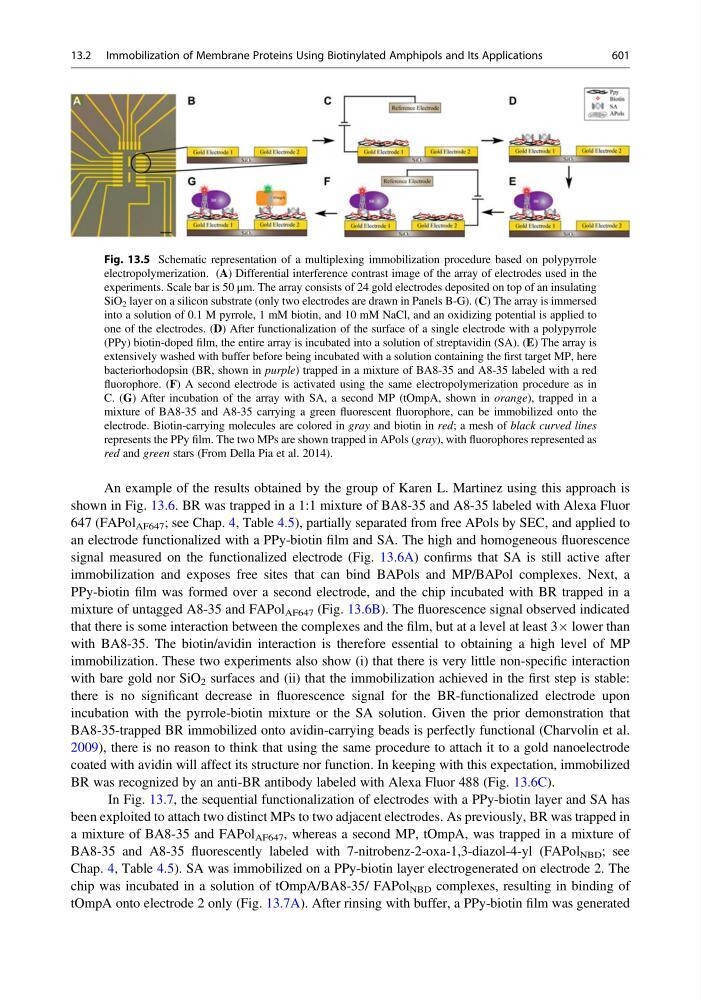
An example of the results obtained by the group of Karen L. Martinez using this approach is
shown in Fig. 13.6. BR was trapped in a 1:1 mixture of BA8-35 and A8-35 labeled with Alexa Fluor
647 (FAPol AF647 ; see Chap. 4, Table 4.5), partially separated from free APols by SEC, and applied to
an electrode functionalized with a PPy-biotin film and SA. The high and homogeneous fluorescence
signal measured on the functionalized electrode (Fig. 13.6A) confirms that SA is still active after
immobilization and exposes free sites that can bind BAPols and MP/BAPol complexes. Next, a
PPy-biotin film was formed over a second electrode, and the chip incubated with BR trapped in a
mixture of untagged A8-35 and FAPol AF647 (Fig. 13.6B). The fluorescence signal observed indicated
that there is some interaction between the complexes and the film, but at a level at least 3Â lower than
with BA8-35. The biotin/avidin interaction is therefore essential to obtaining a high level of MP
immobilization. These two experiments also show (i) that there is very little non-specific interaction
with bare gold nor SiO 2 surfaces and (ii) that the immobilization achieved in the first step is stable:
there is no significant decrease in fluorescence signal for the BR-functionalized electrode upon
incubation with the pyrrole-biotin mixture or the SA solution. Given the prior demonstration that
BA8-35-trapped BR immobilized onto avidin-carrying beads is perfectly functional (Charvolin et al.
2009), there is no reason to think that using the same procedure to attach it to a gold nanoelectrode
coated with avidin will affect its structure nor function. In keeping with this expectation, immobilized
BR was recognized by an anti-BR antibody labeled with Alexa Fluor 488 (Fig. 13.6C).
In Fig. 13.7, the sequential functionalization of electrodes with a PPy-biotin layer and SA has
been exploited to attach two distinct MPs to two adjacent electrodes. As previously, BR was trapped in
a mixture of BA8-35 and FAPol AF647 , whereas a second MP, tOmpA, was trapped in a mixture of
BA8-35 and A8-35 fluorescently labeled with 7-nitrobenz-2-oxa-1,3-diazol-4-yl (FAPol NBD ; see
Chap. 4, Table 4.5). SA was immobilized on a PPy-biotin layer electrogenerated on electrode 2. The
chip was incubated in a solution of tOmpA/BA8-35/ FAPol NBD complexes, resulting in binding of
tOmpA onto electrode 2 only (Fig. 13.7A). After rinsing with buffer, a PPy-biotin film was generated
Fig. 13.5 Schematic representation of a multiplexing immobilization procedure based on polypyrrole
electropolymerization. (A) Differential interference contrast image of the array of electrodes used in the
experiments. Scale bar is 50 μm. The array consists of 24 gold electrodes deposited on top of an insulating
SiO 2 layer on a silicon substrate (only two electrodes are drawn in Panels B-G). (C) The array is immersed
into a solution of 0.1 M pyrrole, 1 mM biotin, and 10 mM NaCl, and an oxidizing potential is applied to
one of the electrodes. (D) After functionalization of the surface of a single electrode with a polypyrrole
(PPy) biotin-doped film, the entire array is incubated into a solution of streptavidin (SA). (E) The array is
extensively washed with buffer before being incubated with a solution containing the first target MP, here
bacteriorhodopsin (BR, shown in purple) trapped in a mixture of BA8-35 and A8-35 labeled with a red
fluorophore. (F) A second electrode is activated using the same electropolymerization procedure as in
C. (G) After incubation of the array with SA, a second MP (tOmpA, shown in orange), trapped in a
mixture of BA8-35 and A8-35 carrying a green fluorescent fluorophore, can be immobilized onto the
electrode. Biotin-carrying molecules are colored in gray and biotin in red; a mesh of black curved lines
represents the PPy film. The two MPs are shown trapped in APols (gray), with fluorophores represented as
red and green stars (From Della Pia et al. 2014).
13.2 Immobilization of Membrane Proteins Using Biotinylated Amphipols and Its Applications
601