

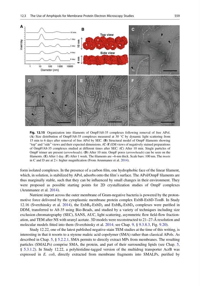
form isolated complexes. In the presence of a carbon film, one hydrophobic face of the linear filament,
which, in solution, is stabilized by APol, adsorbs onto the film’s surface. The APol/OmpF filaments are
thus marginally stable, such that they can be influenced by small changes in their environment. They
were proposed as possible starting points for 2D crystallization studies of OmpF complexes
(Arunmanee et al. 2014).
Nutrient import across the outer membrane of Gram-negative bacteria is powered by the protonmotive force delivered by the cytoplasmic membrane protein complex ExbB-ExbD-TonB. In Study
12.16 (Sverzhinsky et al. 2014), the ExbB 4 -ExbD 2 and ExbB 6 -ExbD 4 complexes were purified in
DDM, transferred to A8-35 using Bio-Beads, and studied by a variety of techniques including size
exclusion chromatography (SEC), SANS, AUC, light scattering, asymmetric flow field-flow fractionation, and TEM after NS with uranyl acetate. 3D models were reconstructed to 21–27-Å resolution and
molecular models fitted into them (Sverzhinsky et al. 2014; see Chap. 9, § 9.3.8.3, Fig. 9.20).
Study 12.22, one of the latest published negative-stain TEM studies at the time of this writing, is
interesting in that it resorts to a styrene maleic acid copolymer (SMA) rather than classical APols. As
described in Chap. 5, § 5.2.2.1, SMA permits to directly extract MPs from membranes. The resulting
particles (SMALPs) comprise SMA, the protein, and part of their surrounding lipids (see Chap. 5,
§ 5.3.1.2). In Study 12.22, a polyhistidine-tagged version of the multidrug transporter AcrB was
expressed in E. coli, directly extracted from membrane fragments into SMALPs, purified by
Fig. 12.15 Organization into filaments of OmpF/A8-35 complexes following removal of free APol.
(A) Size distribution of OmpF/A8-35 complexes measured at 30
C by dynamic light scattering from
15 min to 6 days after removal of free APol by SEC. (B) Structural model of OmpF filaments showing
“top” and “side” views and their expected dimensions. (C–F) EM views of negatively stained preparations
of OmpF/A8-35 complexes studied at different times after SEC: (C) After 10 min. Single particles of
OmpF trimer are present (arrowheads). (D) After 10 min. OmpF pores (arrowheads) can be seen on the
filaments. (E) After 1 day. (F) After 1 week. The filaments are ~6-nm thick. Scale bars: 100 nm. The insets
in C and D are at 2Â higher magnification (From Arunmanee et al. 2014).
12.3 The Use of Amphipols for Membrane Protein Electron Microscopy Studies
559
which, in solution, is stabilized by APol, adsorbs onto the film’s surface. The APol/OmpF filaments are
thus marginally stable, such that they can be influenced by small changes in their environment. They
were proposed as possible starting points for 2D crystallization studies of OmpF complexes
(Arunmanee et al. 2014).
Nutrient import across the outer membrane of Gram-negative bacteria is powered by the protonmotive force delivered by the cytoplasmic membrane protein complex ExbB-ExbD-TonB. In Study
12.16 (Sverzhinsky et al. 2014), the ExbB 4 -ExbD 2 and ExbB 6 -ExbD 4 complexes were purified in
DDM, transferred to A8-35 using Bio-Beads, and studied by a variety of techniques including size
exclusion chromatography (SEC), SANS, AUC, light scattering, asymmetric flow field-flow fractionation, and TEM after NS with uranyl acetate. 3D models were reconstructed to 21–27-Å resolution and
molecular models fitted into them (Sverzhinsky et al. 2014; see Chap. 9, § 9.3.8.3, Fig. 9.20).
Study 12.22, one of the latest published negative-stain TEM studies at the time of this writing, is
interesting in that it resorts to a styrene maleic acid copolymer (SMA) rather than classical APols. As
described in Chap. 5, § 5.2.2.1, SMA permits to directly extract MPs from membranes. The resulting
particles (SMALPs) comprise SMA, the protein, and part of their surrounding lipids (see Chap. 5,
§ 5.3.1.2). In Study 12.22, a polyhistidine-tagged version of the multidrug transporter AcrB was
expressed in E. coli, directly extracted from membrane fragments into SMALPs, purified by
Fig. 12.15 Organization into filaments of OmpF/A8-35 complexes following removal of free APol.
(A) Size distribution of OmpF/A8-35 complexes measured at 30
C by dynamic light scattering from
15 min to 6 days after removal of free APol by SEC. (B) Structural model of OmpF filaments showing
“top” and “side” views and their expected dimensions. (C–F) EM views of negatively stained preparations
of OmpF/A8-35 complexes studied at different times after SEC: (C) After 10 min. Single particles of
OmpF trimer are present (arrowheads). (D) After 10 min. OmpF pores (arrowheads) can be seen on the
filaments. (E) After 1 day. (F) After 1 week. The filaments are ~6-nm thick. Scale bars: 100 nm. The insets
in C and D are at 2Â higher magnification (From Arunmanee et al. 2014).
12.3 The Use of Amphipols for Membrane Protein Electron Microscopy Studies
559