

immobilized-metal affinity chromatography (IMAC), and imaged after staining with uranyl acetate
(Postis et al. 2015). The particles appeared as either “singlets” or “doublets,” the images of which were
separately analyzed. A 3D model of “singlets,” reconstructed to a final resolution of 23 Å, is consistent
with the known X-ray structure of the AcrB trimer (Murakami et al. 2002), with SMA + lipids (SMAL)
forming a belt around the TM region. Class analyses of the doublets show a variety of association
modes, all of them seeming to involve contacts between the SMAL belts of two AcrB trimers. Control
experiments suggest that the formation of doublets occurs in solution and not on the EM grid. The lack
of any preferred doublet interface suggests that particle association occurs non-specifically via the
SMAL belts and does not involve any defined protein/protein interface. These observations suggest
that any SMAL-trapped MP is likely to behave in the same way. Note that, as also observed with
OmpF/A8-35 complexes (Study 12.13; Arunmanee et al. 2014), AcrB/SMAL ones tend to adsorb onto
the carbon film sideways, with their TM surface in contact with the carbon, even though the latter has
been made hydrophilic by glow-discharge.
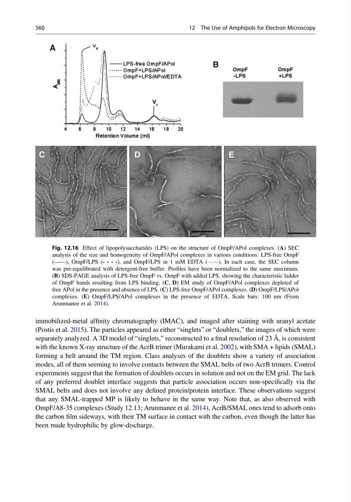
Fig. 12.16 Effect of lipopolysaccharides (LPS) on the structure of OmpF/APol complexes. (A) SEC
analysis of the size and homogeneity of OmpF/APol complexes in various conditions: LPS-free OmpF
(——), OmpF/LPS (- - - -), and OmpF/LPS in 1 mM EDTA (ÁÁÁÁÁÁÁ). In each case, the SEC column
was pre-equilibrated with detergent-free buffer. Profiles have been normalized to the same maximum.
(B) SDS-PAGE analysis of LPS-free OmpF vs. OmpF with added LPS, showing the characteristic ladder
of OmpF bands resulting from LPS binding. (C, D) EM study of OmpF/APol complexes depleted of
free APol in the presence and absence of LPS. (C) LPS-free OmpF/APol complexes. (D) OmpF/LPS/APol
complexes. (E) OmpF/LPS/APol complexes in the presence of EDTA. Scale bars: 100 nm (From
Arunmanee et al. 2014).
560
12 The Use of Amphipols for Electron Microscopy
(Postis et al. 2015). The particles appeared as either “singlets” or “doublets,” the images of which were
separately analyzed. A 3D model of “singlets,” reconstructed to a final resolution of 23 Å, is consistent
with the known X-ray structure of the AcrB trimer (Murakami et al. 2002), with SMA + lipids (SMAL)
forming a belt around the TM region. Class analyses of the doublets show a variety of association
modes, all of them seeming to involve contacts between the SMAL belts of two AcrB trimers. Control
experiments suggest that the formation of doublets occurs in solution and not on the EM grid. The lack
of any preferred doublet interface suggests that particle association occurs non-specifically via the
SMAL belts and does not involve any defined protein/protein interface. These observations suggest
that any SMAL-trapped MP is likely to behave in the same way. Note that, as also observed with
OmpF/A8-35 complexes (Study 12.13; Arunmanee et al. 2014), AcrB/SMAL ones tend to adsorb onto
the carbon film sideways, with their TM surface in contact with the carbon, even though the latter has
been made hydrophilic by glow-discharge.
Fig. 12.16 Effect of lipopolysaccharides (LPS) on the structure of OmpF/APol complexes. (A) SEC
analysis of the size and homogeneity of OmpF/APol complexes in various conditions: LPS-free OmpF
(——), OmpF/LPS (- - - -), and OmpF/LPS in 1 mM EDTA (ÁÁÁÁÁÁÁ). In each case, the SEC column
was pre-equilibrated with detergent-free buffer. Profiles have been normalized to the same maximum.
(B) SDS-PAGE analysis of LPS-free OmpF vs. OmpF with added LPS, showing the characteristic ladder
of OmpF bands resulting from LPS binding. (C, D) EM study of OmpF/APol complexes depleted of
free APol in the presence and absence of LPS. (C) LPS-free OmpF/APol complexes. (D) OmpF/LPS/APol
complexes. (E) OmpF/LPS/APol complexes in the presence of EDTA. Scale bars: 100 nm (From
Arunmanee et al. 2014).
560
12 The Use of Amphipols for Electron Microscopy