

Shao et al. (2016), Abeyrathne and Grigorieff (2017), Bausewein et al. (2017), Chiu et al. (2017), He
et al. (2017), and Zhou et al. (2017). For a recent review on TRP cryo-EM studies, see Madej and
Ziegler 2018.
The negative-stain study of TRPA1 by Vera Y. Moiseenkova-Bell and colleagues (Cvetkov et al.
2011; Huynh et al. 2014a, b; Study 12.6 in Table 12.1) was the first to introduce the use of APols for
EM studies of the TRP channel family and was to have a rich posterity. Mouse TRPA1 was expressed
in Saccharomyces cerevisiae, solubilized and purified in Fos-Choline-12 (FC12), and transferred to
A8-35 by dilution under the CMC of FC12 and dialysis in the presence of Bio-Beads. Under these
conditions, most of the free A8-35 must have remained present in the samples. The TRPA1/A8-35
complexes were adsorbed onto carbon film-coated copper grids, washed with pure water, and stained
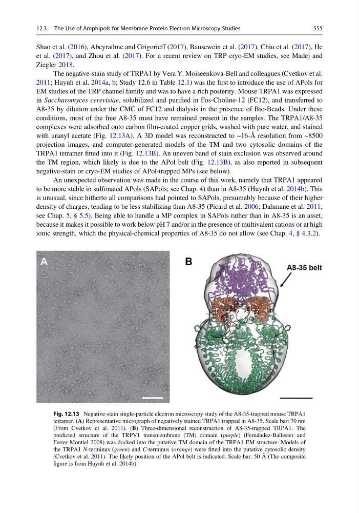
with uranyl acetate (Fig. 12.13A). A 3D model was reconstructed to ~16-Å resolution from ~8500
projection images, and computer-generated models of the TM and two cytosolic domains of the
TRPA1 tetramer fitted into it (Fig. 12.13B). An uneven band of stain exclusion was observed around
the TM region, which likely is due to the APol belt (Fig. 12.13B), as also reported in subsequent
negative-stain or cryo-EM studies of APol-trapped MPs (see below).
An unexpected observation was made in the course of this work, namely that TRPA1 appeared
to be more stable in sulfonated APols (SAPols; see Chap. 4) than in A8-35 (Huynh et al. 2014b). This
is unusual, since hitherto all comparisons had pointed to SAPols, presumably because of their higher
density of charges, tending to be less stabilizing than A8-35 (Picard et al. 2006; Dahmane et al. 2011;
see Chap. 5, § 5.5). Being able to handle a MP complex in SAPols rather than in A8-35 is an asset,
because it makes it possible to work below pH 7 and/or in the presence of multivalent cations or at high
ionic strength, which the physical-chemical properties of A8-35 do not allow (see Chap. 4, § 4.3.2).
Fig. 12.13 Negative-stain single-particle electron microscopy study of the A8-35-trapped mouse TRPA1
tetramer. (A) Representative micrograph of negatively stained TRPA1 trapped in A8-35. Scale bar: 70 nm
(From Cvetkov et al. 2011). (B) Three-dimensional reconstruction of A8-35-trapped TRPA1. The
predicted structure of the TRPV1 transmembrane (TM) domain (purple) (Fernández-Ballester and
Ferrer-Montiel 2008) was docked into the putative TM domain of the TRPA1 EM structure. Models of
the TRPA1 N-terminus (green) and C-terminus (orange) were fitted into the putative cytosolic density
(Cvetkov et al. 2011). The likely position of the APol belt is indicated. Scale bar: 50 Å (The composite
figure is from Huynh et al. 2014b).
12.3 The Use of Amphipols for Membrane Protein Electron Microscopy Studies
555
et al. (2017), and Zhou et al. (2017). For a recent review on TRP cryo-EM studies, see Madej and
Ziegler 2018.
The negative-stain study of TRPA1 by Vera Y. Moiseenkova-Bell and colleagues (Cvetkov et al.
2011; Huynh et al. 2014a, b; Study 12.6 in Table 12.1) was the first to introduce the use of APols for
EM studies of the TRP channel family and was to have a rich posterity. Mouse TRPA1 was expressed
in Saccharomyces cerevisiae, solubilized and purified in Fos-Choline-12 (FC12), and transferred to
A8-35 by dilution under the CMC of FC12 and dialysis in the presence of Bio-Beads. Under these
conditions, most of the free A8-35 must have remained present in the samples. The TRPA1/A8-35
complexes were adsorbed onto carbon film-coated copper grids, washed with pure water, and stained
with uranyl acetate (Fig. 12.13A). A 3D model was reconstructed to ~16-Å resolution from ~8500
projection images, and computer-generated models of the TM and two cytosolic domains of the
TRPA1 tetramer fitted into it (Fig. 12.13B). An uneven band of stain exclusion was observed around
the TM region, which likely is due to the APol belt (Fig. 12.13B), as also reported in subsequent
negative-stain or cryo-EM studies of APol-trapped MPs (see below).
An unexpected observation was made in the course of this work, namely that TRPA1 appeared
to be more stable in sulfonated APols (SAPols; see Chap. 4) than in A8-35 (Huynh et al. 2014b). This
is unusual, since hitherto all comparisons had pointed to SAPols, presumably because of their higher
density of charges, tending to be less stabilizing than A8-35 (Picard et al. 2006; Dahmane et al. 2011;
see Chap. 5, § 5.5). Being able to handle a MP complex in SAPols rather than in A8-35 is an asset,
because it makes it possible to work below pH 7 and/or in the presence of multivalent cations or at high
ionic strength, which the physical-chemical properties of A8-35 do not allow (see Chap. 4, § 4.3.2).
Fig. 12.13 Negative-stain single-particle electron microscopy study of the A8-35-trapped mouse TRPA1
tetramer. (A) Representative micrograph of negatively stained TRPA1 trapped in A8-35. Scale bar: 70 nm
(From Cvetkov et al. 2011). (B) Three-dimensional reconstruction of A8-35-trapped TRPA1. The
predicted structure of the TRPV1 transmembrane (TM) domain (purple) (Fernández-Ballester and
Ferrer-Montiel 2008) was docked into the putative TM domain of the TRPA1 EM structure. Models of
the TRPA1 N-terminus (green) and C-terminus (orange) were fitted into the putative cytosolic density
(Cvetkov et al. 2011). The likely position of the APol belt is indicated. Scale bar: 50 Å (The composite
figure is from Huynh et al. 2014b).
12.3 The Use of Amphipols for Membrane Protein Electron Microscopy Studies
555