

that may be of general interest to microscopists working on different systems rather than on the
biological relevance of each specific study.
We will consider first recent studies carried on negatively stained samples of APol-trapped MPs.
12.3.2 Recent Studies of Negatively Stained MP/APol Complexes (2011–Present)
The regulated channels known under the rather awkward name of “transient receptor potential” (TRP)
channels form a large family of tetrameric, Ca
2+ -permeable, nonselective cation channels, which are
implicated in a broad range of cellular processes including pain and temperature sensing, neuronal
development, angiogenesis, cardiac and pulmonary function, and cancer (reviewed in Venkatachalam
and Montell 2007). They play key roles in such cellular processes as vision, hearing, touch, pain, and
thermo- and mechanosensory transduction and, as such, are important pharmacological targets.
Mammalian TRP channels share ~20% sequence similarity and are distributed into six subfamilies:
TRPC (canonical), TRPV (vanilloid), TRPA (ankyrin), TRPM (melastatin), TRPP (polycystin), and
TRPML (mucolipin). They have resisted 3D crystallization but appear to be particularly amenable to
single-particle EM studies, particularly as APol-trapped complexes. Seven such studies have been
published to date, one in negative stain, that of TRPA1 (Cvetkov et al. 2011; Huynh et al. 2014b; Study
12.6 in Table 12.1), and six by cryo-EM, those of TRPV1 (Cao et al. 2013b; Liao et al. 2013, 2014;
Study 12.9), TRPM1 (Agosto et al. 2014; Study 12.12), TRPV2 (Fan et al. 2014; Study 12.18), TRPA1
(Paulsen et al. 2015; Study 12.21), TRPV2 again (Zubcevic et al. 2016; Study 12.29), and polycystin2 (Wilkes et al. 2017; Study 12.33). To the studies listed in Table 12.1 and discussed below should be
added seven further relevant studies, which were published or located too late to be integrated in the
table and the discussion, namely those by Spear et al. (2015), Wu et al. (2015), Zhang et al. (2015),
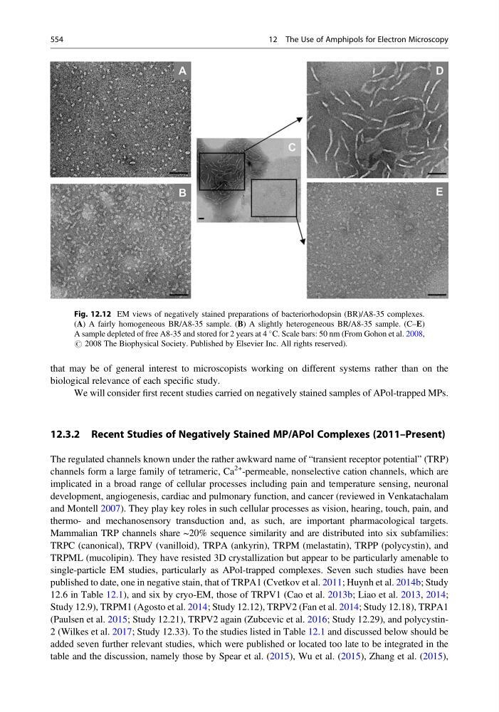
Fig. 12.12 EM views of negatively stained preparations of bacteriorhodopsin (BR)/A8-35 complexes.
(A) A fairly homogeneous BR/A8-35 sample. (B) A slightly heterogeneous BR/A8-35 sample. (C–E)
A sample depleted of free A8-35 and stored for 2 years at 4
C. Scale bars: 50 nm (From Gohon et al. 2008,
# 2008 The Biophysical Society. Published by Elsevier Inc. All rights reserved).
554
12 The Use of Amphipols for Electron Microscopy
biological relevance of each specific study.
We will consider first recent studies carried on negatively stained samples of APol-trapped MPs.
12.3.2 Recent Studies of Negatively Stained MP/APol Complexes (2011–Present)
The regulated channels known under the rather awkward name of “transient receptor potential” (TRP)
channels form a large family of tetrameric, Ca
2+ -permeable, nonselective cation channels, which are
implicated in a broad range of cellular processes including pain and temperature sensing, neuronal
development, angiogenesis, cardiac and pulmonary function, and cancer (reviewed in Venkatachalam
and Montell 2007). They play key roles in such cellular processes as vision, hearing, touch, pain, and
thermo- and mechanosensory transduction and, as such, are important pharmacological targets.
Mammalian TRP channels share ~20% sequence similarity and are distributed into six subfamilies:
TRPC (canonical), TRPV (vanilloid), TRPA (ankyrin), TRPM (melastatin), TRPP (polycystin), and
TRPML (mucolipin). They have resisted 3D crystallization but appear to be particularly amenable to
single-particle EM studies, particularly as APol-trapped complexes. Seven such studies have been
published to date, one in negative stain, that of TRPA1 (Cvetkov et al. 2011; Huynh et al. 2014b; Study
12.6 in Table 12.1), and six by cryo-EM, those of TRPV1 (Cao et al. 2013b; Liao et al. 2013, 2014;
Study 12.9), TRPM1 (Agosto et al. 2014; Study 12.12), TRPV2 (Fan et al. 2014; Study 12.18), TRPA1
(Paulsen et al. 2015; Study 12.21), TRPV2 again (Zubcevic et al. 2016; Study 12.29), and polycystin2 (Wilkes et al. 2017; Study 12.33). To the studies listed in Table 12.1 and discussed below should be
added seven further relevant studies, which were published or located too late to be integrated in the
table and the discussion, namely those by Spear et al. (2015), Wu et al. (2015), Zhang et al. (2015),
Fig. 12.12 EM views of negatively stained preparations of bacteriorhodopsin (BR)/A8-35 complexes.
(A) A fairly homogeneous BR/A8-35 sample. (B) A slightly heterogeneous BR/A8-35 sample. (C–E)
A sample depleted of free A8-35 and stored for 2 years at 4
C. Scale bars: 50 nm (From Gohon et al. 2008,
# 2008 The Biophysical Society. Published by Elsevier Inc. All rights reserved).
554
12 The Use of Amphipols for Electron Microscopy