

Fig. 12.12A, B, respectively. The diameter of individual particles was ~6.3 Æ 0.8 nm, with a rather
narrow distribution. The distribution of lengths of the particles presented a first maximum at ~8 nm,
corresponding to the major, globular species (monomeric BR/A8-35 complexes), consistent with
SANS and AUC data, followed by maxima at 13, 19, 25, and 31 nm for the more elongated particles
present in heterogeneous preparations. These views suggested that BR/A8-35 monomers have a
propensity to auto-assemble in a linear way.
This was spectacularly confirmed by observations on a sample of BR/A8-35 complexes depleted
from free APol that had been kept for 2 years at 4
C (A8-35-trapped BR is pretty stable; see Chap. 5).
This preparation was found to comprise a mixture of monomers and small oligomers, similar to those
observed in fresh preparations (Fig. 12.12C, E), but also much longer filaments (Fig. 12.12D). The
filaments showed a fairly regular longitudinal segmentation (Fig. 12.12D). The length of the segments
corresponds roughly to tetra- or pentamers of the globular species. Because the stain formed rather
thick puddles in filament-rich regions (Fig. 12.12C), it is difficult to decide from these micrographs
whether the segmented appearance results from the lengthwise association of smaller oligomers or
from the filaments adopting a helical structure that rises periodically above the carbon film. Related
observations were to be done later on preparations of OmpF/A8-35 complexes (see below, Study
12.13), as well as on PgP/SMAL ones (Study 12.17), and will be commented upon below (§ 12.3.4.2).
Study 12.4 closes what could be termed the prehistory of APol-based EM studies. From then on,
APols and their relatives will be used more and more with biological objectives in mind, with highly
rewarding results, and technological considerations will fade into the background. This second period
opens with the cryo-EM study of the A8-35-trapped mitochondrial respirasome (Althoff et al. 2011),
moving toward higher and higher resolutions thanks to the technological progress of cryo-EM
summarized in § 12.2.3, with, in parallel, the development of negative-stain works. From 2011 on,
APols have been considered more and more as just another surfactant to be tested when launching an
EM study. Whereas the interest in the biological conclusions derived from studying MP/APol
complexes by EM grew, scant attention was paid to the peculiarities of APol physical-chemical
properties and the technical aspects of their use. Our description of the two dozen studies published
in this second period will therefore remain rather sketchy, with the focus placed on those observations
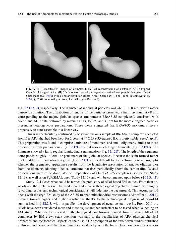
Fig. 12.11 Reconstructed images of Complex I. (A) 3D reconstruction of unstained A8-35-trapped
Complex I imaged in ice. (B) 3D reconstruction of the negatively stained complex in detergent (From
Guénebaut et al. 1998) with a similar resolution cutoff (6 nm). Scale bar: 10 nm (From Flötenmeyer et al.
2007, # 2007 John Wiley & Sons, Inc. All Rights Reserved).
12.3 The Use of Amphipols for Membrane Protein Electron Microscopy Studies
553
narrow distribution. The distribution of lengths of the particles presented a first maximum at ~8 nm,
corresponding to the major, globular species (monomeric BR/A8-35 complexes), consistent with
SANS and AUC data, followed by maxima at 13, 19, 25, and 31 nm for the more elongated particles
present in heterogeneous preparations. These views suggested that BR/A8-35 monomers have a
propensity to auto-assemble in a linear way.
This was spectacularly confirmed by observations on a sample of BR/A8-35 complexes depleted
from free APol that had been kept for 2 years at 4
C (A8-35-trapped BR is pretty stable; see Chap. 5).
This preparation was found to comprise a mixture of monomers and small oligomers, similar to those
observed in fresh preparations (Fig. 12.12C, E), but also much longer filaments (Fig. 12.12D). The
filaments showed a fairly regular longitudinal segmentation (Fig. 12.12D). The length of the segments
corresponds roughly to tetra- or pentamers of the globular species. Because the stain formed rather
thick puddles in filament-rich regions (Fig. 12.12C), it is difficult to decide from these micrographs
whether the segmented appearance results from the lengthwise association of smaller oligomers or
from the filaments adopting a helical structure that rises periodically above the carbon film. Related
observations were to be done later on preparations of OmpF/A8-35 complexes (see below, Study
12.13), as well as on PgP/SMAL ones (Study 12.17), and will be commented upon below (§ 12.3.4.2).
Study 12.4 closes what could be termed the prehistory of APol-based EM studies. From then on,
APols and their relatives will be used more and more with biological objectives in mind, with highly
rewarding results, and technological considerations will fade into the background. This second period
opens with the cryo-EM study of the A8-35-trapped mitochondrial respirasome (Althoff et al. 2011),
moving toward higher and higher resolutions thanks to the technological progress of cryo-EM
summarized in § 12.2.3, with, in parallel, the development of negative-stain works. From 2011 on,
APols have been considered more and more as just another surfactant to be tested when launching an
EM study. Whereas the interest in the biological conclusions derived from studying MP/APol
complexes by EM grew, scant attention was paid to the peculiarities of APol physical-chemical
properties and the technical aspects of their use. Our description of the two dozen studies published
in this second period will therefore remain rather sketchy, with the focus placed on those observations
Fig. 12.11 Reconstructed images of Complex I. (A) 3D reconstruction of unstained A8-35-trapped
Complex I imaged in ice. (B) 3D reconstruction of the negatively stained complex in detergent (From
Guénebaut et al. 1998) with a similar resolution cutoff (6 nm). Scale bar: 10 nm (From Flötenmeyer et al.
2007, # 2007 John Wiley & Sons, Inc. All Rights Reserved).
12.3 The Use of Amphipols for Membrane Protein Electron Microscopy Studies
553