

A 3D reconstruction by this method was carried out using the full data set of 1200 particles, with,
as a reference to determine the tilt angles for the individual images, >3000 projections of the 2.8 nm
resolution 3D reconstruction made from negatively stained Complex I (Guénebaut et al. 1998). These
were cross-correlated with the single-particle cryo-EM images to obtain a set of projections at known
Euler angles, which were then combined by weighted back-projection to give a 3D model
(Fig. 12.11A). The resolution estimated by Fourier shell correlation (Saxton and Baumeister 1982)
for reconstructions from two half data sets was 5.8 nm. A low-pass filtered model of the negatively
stained reconstruction (Guénebaut et al. 1998) at 6 nm was calculated for comparison (Fig. 12.11B).
The low resolution of the 3D reconstruction obtained can be explained partly by the small
number of particles sampled. Another factor may have been the inherent flexibility of the structure of
Complex I. A hinge movement of the two arms relative to each other, which was not taken into account
when carrying out the reconstruction from the cryo-EM images, may have resulted in some blurring.
The membrane arm of Complex I, which has the greater contrast, tends to dominate the alignment
procedure. The result is that the TM arm is similar in shape and size to that observed in negative stain,
whereas the cytoplasmic arm is reduced (Fig. 12.11). However, why this problem should have arisen in
Study 12.3 and not in more recent ones (Zickermann et al. 2009; Vinothkumar et al. 2014) is not clear.
The fact that the use of APols resulted in ice films with a more uniform thickness and therefore
a more even distribution of particles compared to that observed in the presence of detergents is a
particularly interesting observation, whose origin will be discussed below (§ 12.3.4.2).
The period under consideration closes with Study 12.4, in which BR/A8-35 complexes were
examined by negative staining in the frame of a broader study of their composition and properties
(Gohon et al. 2008). This study identified some of the factors that could lead to heterogeneous
preparations, such as (i) problems arising during the synthesis of the polymers, making them more
hydrophobic than had been aimed for (Gohon et al. 2006, 2008; see Chap. 4), and (ii) removal of free
APol (Zoonens et al. 2007; see Chap. 5). EM views of preparations that had been identified as
homogeneous or heterogeneous by small-angle neutron scattering (SANS) and AUC are shown in
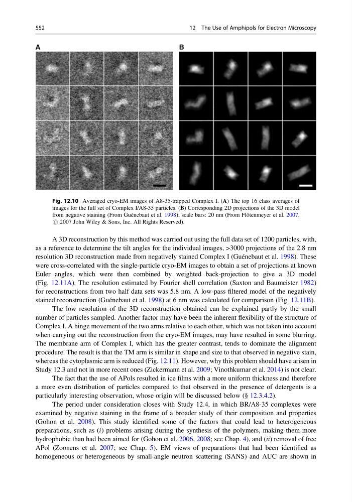
Fig. 12.10 Averaged cryo-EM images of A8-35-trapped Complex I. (A) The top 16 class averages of
images for the full set of Complex I/A8-35 particles. (B) Corresponding 2D projections of the 3D model
from negative staining (From Guénebaut et al. 1998); scale bars: 20 nm (From Flötenmeyer et al. 2007,
# 2007 John Wiley & Sons, Inc. All Rights Reserved).
552
12 The Use of Amphipols for Electron Microscopy
as a reference to determine the tilt angles for the individual images, >3000 projections of the 2.8 nm
resolution 3D reconstruction made from negatively stained Complex I (Guénebaut et al. 1998). These
were cross-correlated with the single-particle cryo-EM images to obtain a set of projections at known
Euler angles, which were then combined by weighted back-projection to give a 3D model
(Fig. 12.11A). The resolution estimated by Fourier shell correlation (Saxton and Baumeister 1982)
for reconstructions from two half data sets was 5.8 nm. A low-pass filtered model of the negatively
stained reconstruction (Guénebaut et al. 1998) at 6 nm was calculated for comparison (Fig. 12.11B).
The low resolution of the 3D reconstruction obtained can be explained partly by the small
number of particles sampled. Another factor may have been the inherent flexibility of the structure of
Complex I. A hinge movement of the two arms relative to each other, which was not taken into account
when carrying out the reconstruction from the cryo-EM images, may have resulted in some blurring.
The membrane arm of Complex I, which has the greater contrast, tends to dominate the alignment
procedure. The result is that the TM arm is similar in shape and size to that observed in negative stain,
whereas the cytoplasmic arm is reduced (Fig. 12.11). However, why this problem should have arisen in
Study 12.3 and not in more recent ones (Zickermann et al. 2009; Vinothkumar et al. 2014) is not clear.
The fact that the use of APols resulted in ice films with a more uniform thickness and therefore
a more even distribution of particles compared to that observed in the presence of detergents is a
particularly interesting observation, whose origin will be discussed below (§ 12.3.4.2).
The period under consideration closes with Study 12.4, in which BR/A8-35 complexes were
examined by negative staining in the frame of a broader study of their composition and properties
(Gohon et al. 2008). This study identified some of the factors that could lead to heterogeneous
preparations, such as (i) problems arising during the synthesis of the polymers, making them more
hydrophobic than had been aimed for (Gohon et al. 2006, 2008; see Chap. 4), and (ii) removal of free
APol (Zoonens et al. 2007; see Chap. 5). EM views of preparations that had been identified as
homogeneous or heterogeneous by small-angle neutron scattering (SANS) and AUC are shown in
Fig. 12.10 Averaged cryo-EM images of A8-35-trapped Complex I. (A) The top 16 class averages of
images for the full set of Complex I/A8-35 particles. (B) Corresponding 2D projections of the 3D model
from negative staining (From Guénebaut et al. 1998); scale bars: 20 nm (From Flötenmeyer et al. 2007,
# 2007 John Wiley & Sons, Inc. All Rights Reserved).
552
12 The Use of Amphipols for Electron Microscopy