

In the same review (Huynh et al. 2014b), it is noted that another TRP channel, TRPV2, is
stabilized in its tetrameric state upon being transferred from DDM to A8-35, whereas exchange to
SAPols promotes the aggregation of TRPV2 into higher oligomeric structures. This initiates a theme
that will run throughout the rest of this chapter: it is currently impossible to predict which APols, or
other surfactants, will turn out to provide the best preparations for an EM study. It is therefore
advisable, at the onset of such a study, to screen several of them to identify the molecule and conditions
that yield the best stability, dispersity, and homogeneity of the particles. In the case of TRPV2, it
turned out that it is one of the MNG detergents that yielded the most homogeneous preparations as
determined by SEC, Blue-Native gel, and NS EM, and it is in this surfactant that the cryo-EM study
was performed, reaching a resolution of 13.6 Å (Huynh et al. 2014a). Such comparisons, however,
should be taken with caution, as there is a degree of chance involved: once satisfactory images have
been obtained with one surfactant, investigators will not necessarily invest months of work examining
whether an apparently inferior one would not yield even better data under slightly different conditions,
so the order in which surfactants and experimental conditions are tested plays a role in the final choice.
TRPV2 is a good example: in the same year one group recommended decyl MNG over A8-35, another
reported having reached essentially the same resolution (~15 Å) in A8-35 (Fan et al. 2014; Study
12.18), and high-resolution structures were finally obtained both in A8-35 (to ~4 Å) (Zubcevic et al.
2016; Study 12.29) and in decyl MNG (to ~5 Å) (Huynh et al. 2016). In the case of the
mechanosensitive channel Piezo1, NS images were recorded in C 12 E 10 , C 12 E 8 , and A8-35. C 12 E 10
was chosen for cryo-EM analysis because it produced slightly better micrographs (Ge et al. 2015). The
N-type rotary ATPase from Burkholderia pseudomallei yielded much better images in lauryldimethylaminoxide than in A8-35 (Schulz et al. 2017), whereas better images of the mechanotransduction
channel NOMPC were obtained in nanodiscs than in A8-35 (Jin et al. 2017).
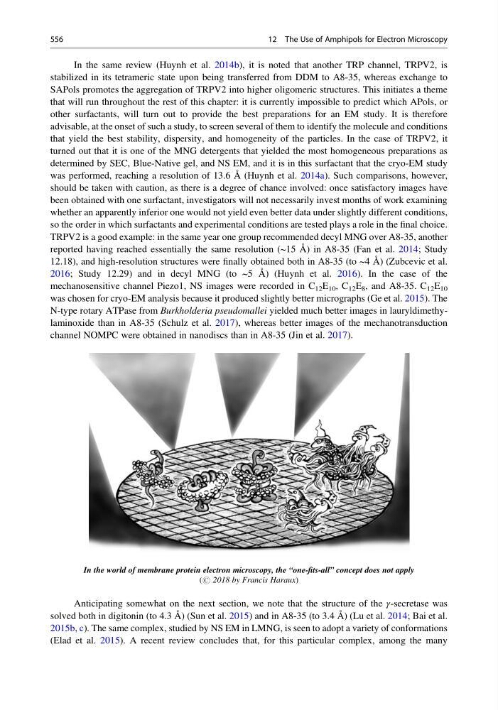
In the world of membrane protein electron microscopy, the “one-fits-all” concept does not apply
(# 2018 by Francis Haraux)
Anticipating somewhat on the next section, we note that the structure of the γ-secretase was
solved both in digitonin (to 4.3 Å) (Sun et al. 2015) and in A8-35 (to 3.4 Å) (Lu et al. 2014; Bai et al.
2015b, c). The same complex, studied by NS EM in LMNG, is seen to adopt a variety of conformations
(Elad et al. 2015). A recent review concludes that, for this particular complex, among the many
556
12 The Use of Amphipols for Electron Microscopy
stabilized in its tetrameric state upon being transferred from DDM to A8-35, whereas exchange to
SAPols promotes the aggregation of TRPV2 into higher oligomeric structures. This initiates a theme
that will run throughout the rest of this chapter: it is currently impossible to predict which APols, or
other surfactants, will turn out to provide the best preparations for an EM study. It is therefore
advisable, at the onset of such a study, to screen several of them to identify the molecule and conditions
that yield the best stability, dispersity, and homogeneity of the particles. In the case of TRPV2, it
turned out that it is one of the MNG detergents that yielded the most homogeneous preparations as
determined by SEC, Blue-Native gel, and NS EM, and it is in this surfactant that the cryo-EM study
was performed, reaching a resolution of 13.6 Å (Huynh et al. 2014a). Such comparisons, however,
should be taken with caution, as there is a degree of chance involved: once satisfactory images have
been obtained with one surfactant, investigators will not necessarily invest months of work examining
whether an apparently inferior one would not yield even better data under slightly different conditions,
so the order in which surfactants and experimental conditions are tested plays a role in the final choice.
TRPV2 is a good example: in the same year one group recommended decyl MNG over A8-35, another
reported having reached essentially the same resolution (~15 Å) in A8-35 (Fan et al. 2014; Study
12.18), and high-resolution structures were finally obtained both in A8-35 (to ~4 Å) (Zubcevic et al.
2016; Study 12.29) and in decyl MNG (to ~5 Å) (Huynh et al. 2016). In the case of the
mechanosensitive channel Piezo1, NS images were recorded in C 12 E 10 , C 12 E 8 , and A8-35. C 12 E 10
was chosen for cryo-EM analysis because it produced slightly better micrographs (Ge et al. 2015). The
N-type rotary ATPase from Burkholderia pseudomallei yielded much better images in lauryldimethylaminoxide than in A8-35 (Schulz et al. 2017), whereas better images of the mechanotransduction
channel NOMPC were obtained in nanodiscs than in A8-35 (Jin et al. 2017).
In the world of membrane protein electron microscopy, the “one-fits-all” concept does not apply
(# 2018 by Francis Haraux)
Anticipating somewhat on the next section, we note that the structure of the γ-secretase was
solved both in digitonin (to 4.3 Å) (Sun et al. 2015) and in A8-35 (to 3.4 Å) (Lu et al. 2014; Bai et al.
2015b, c). The same complex, studied by NS EM in LMNG, is seen to adopt a variety of conformations
(Elad et al. 2015). A recent review concludes that, for this particular complex, among the many
556
12 The Use of Amphipols for Electron Microscopy