

Study
Membrane protein
(MW)
Amphipol
EM
methods
Resolution
(Å)
Main observations relevant to the use of amphipols for EM
References
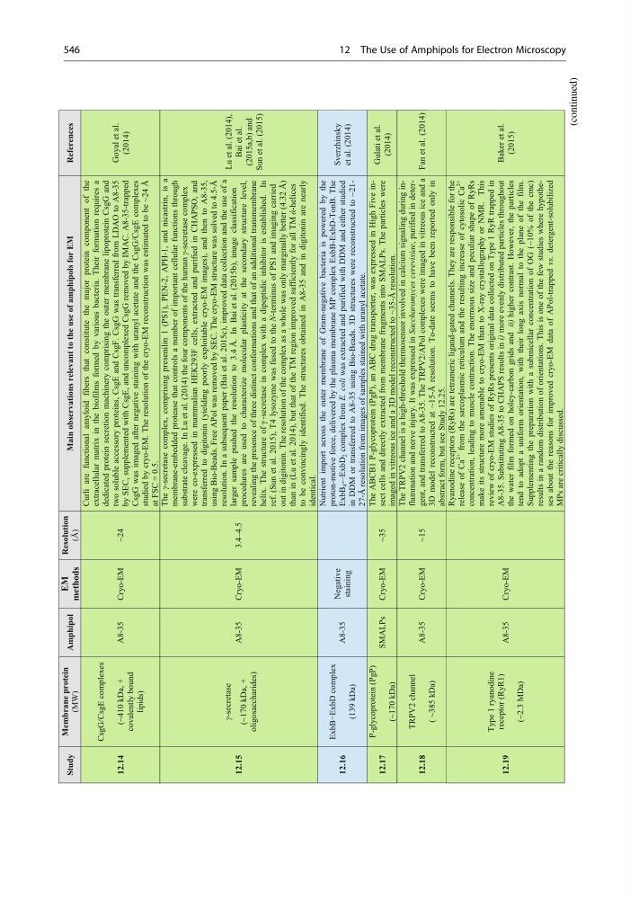
12.14
CsgG/CsgE complexes
(~410 kDa, +
covalently bound
lipids)
A8-35
Cryo-EM
~24
Curli are functional amyloid fibers that constitute the major protein component of the
extracellular matrix in the biofilms formed by various bacteria. Their formation requires a
dedicated protein secretion machinery comprising the outer membrane lipoprotein CsgG and
two soluble accessory proteins, CsgE and CsgF. CsgG was transferred from LDAO to A8-35
by SEC, supplemented with CsgE, and uncomplexed CsgG removed by IMAC. A8-35-trapped
CsgG was imaged after negative staining with uranyl acetate and the CsgG/CsgE complexes
studied by cryo-EM. The resolution of the cryo-EM reconstruction was estimated to be ~24 Å
at FSC = 0.5.
Goyal et al.
(2014)
12.15
(~170 kDa, +
oligosaccharides)
A8-35
Cryo-EM
3.4–4.5
The γ-secretase complex, comprising presenilin 1 (PS1), PEN-2, APH-1, and nicastrin, is a
membrane-embedded protease that controls a number of important cellular functions through
substrate cleavage. In Lu et al. (2014) the four components of the human γ-secretase complex
were co-expressed in mammalian HEK293F cells, extracted and purified in CHAPSO, and
transferred to digitonin (yielding poorly exploitable cryo-EM images), and then to A8-35,
using Bio-Beads. Free APol was removed by SEC. The cryo-EM structure was solved to 4.5-Å
resolution. In a subsequent paper (Bai et al. 2015c), improved data collection and the use of a
larger sample pushed the resolution to 3.4 Å. In Bai et al. (2015b), image classification
procedures are used to characterize molecular plasticity at the secondary structure level,
revealing the presence of three distinct conformations and that of an additional transmembrane
helix. The structure of γ -secretase in complex with a dipeptidic inhibitor is established. In
ref. (Sun et al. 2015), T4 lysozyme was fused to the N-terminus of PS1 and imaging carried
out in digitonin. The resolution of the complex as a whole was only marginally better (4.32 Å)
than in (Lu et al. 2014), but that of the TM region improved sufficiently for all TM ά-helices
to be convincingly identified. The structures obtained in A8-35 and in digitonin are nearly
identical.
Lu et al. (2014),
Bai et al.
(2015a,b) and
Sun et al. (2015)
12.16
ExbB−ExbD complex
(139 kDa)
A8-35
Negative
staining
Nutrient import across the outer membrane of Gram-negative bacteria is powered by the
proton-motive force, delivered by the plasma membrane MP complex ExbB-ExbD-TonB. The
ExbB
4 -–ExbD
2 complex from E. coli was extracted and purified with DDM and either studied
in DDM or transferred to A8-35 using Bio-Beads. 3D structures were reconstructed to ~2127-Å resolution from images of samples stained with uranyl acetate.
12.17
P-glycoprotein (PgP)
(~170 kDa)
SMALPs
Cryo-EM
~35
The ABCB1 P-glycoprotein (PgP), an ABC drug transporter, was expressed in High Five insect cells and directly extracted from membrane fragments into SMALPs. The particles were
imaged in vitreous ice and a 3D model reconstructed to ~35-Å resolution.
12.18
TRPV2 channel
( ~385 kDa)
A8-35
Cryo-EM
~15
The TRPV2 channel is a high-threshold thermosensor involved in calcium signaling during inflammation and nerve injury. It was expressed in Saccharomyces cerevisiae, purified in detergent, and transferred to A8-35. The TRPV2/APol complexes were imaged in vitreous ice and a
3D model reconstructed at ~15-Å resolution. To-date seems to have been reported only in
abstract form, but see Study 12.25.
Fan et al. (2014)
γ-secretase
12.19
Type 1 ryanodine
receptor (RyR1)
(~2.3 MDa)
A8-35
Cryo-EM
Ryanodine receptors (RyRs) are tetrameric ligand-gated channels. They are responsible for the
release of Ca
2+
from the sarcoplasmic reticulum and the resulting increase of cytosolic Ca
2+
concentration, leading to muscle contraction. The enormous size and peculiar shape of RyRs
make its structure more amenable to cryo-EM than to X-ray crystallography or NMR. This
review of cryo-EM studies of RyRs presents original data collected on Type 1 RyR trapped in
A8-35. Substituting A8-35 to CHAPS results in i) more evenly distributed particles throughout
the water film formed on holey-carbon grids and ii) higher contrast. However, the particles
tend to adopt a uniform orientation, with their long axis normal to the plane of the film.
Supplementing the preparation with a submicellar concentration of OG (~10% of the cmc)
results in a random distribution of orientations. This is one of the few studies where hypotheses about the reasons for improved cryo-EM data of APol-trapped vs. detergent-solubilized
MPs are critically discussed.
Gulati et al.
(2014)
Baker et al.
(2015)
Sverzhinsky
et al. (2014)
(continued)
546
12 The Use of Amphipols for Electron Microscopy
Membrane protein
(MW)
Amphipol
EM
methods
Resolution
(Å)
Main observations relevant to the use of amphipols for EM
References
12.14
CsgG/CsgE complexes
(~410 kDa, +
covalently bound
lipids)
A8-35
Cryo-EM
~24
Curli are functional amyloid fibers that constitute the major protein component of the
extracellular matrix in the biofilms formed by various bacteria. Their formation requires a
dedicated protein secretion machinery comprising the outer membrane lipoprotein CsgG and
two soluble accessory proteins, CsgE and CsgF. CsgG was transferred from LDAO to A8-35
by SEC, supplemented with CsgE, and uncomplexed CsgG removed by IMAC. A8-35-trapped
CsgG was imaged after negative staining with uranyl acetate and the CsgG/CsgE complexes
studied by cryo-EM. The resolution of the cryo-EM reconstruction was estimated to be ~24 Å
at FSC = 0.5.
Goyal et al.
(2014)
12.15
(~170 kDa, +
oligosaccharides)
A8-35
Cryo-EM
3.4–4.5
The γ-secretase complex, comprising presenilin 1 (PS1), PEN-2, APH-1, and nicastrin, is a
membrane-embedded protease that controls a number of important cellular functions through
substrate cleavage. In Lu et al. (2014) the four components of the human γ-secretase complex
were co-expressed in mammalian HEK293F cells, extracted and purified in CHAPSO, and
transferred to digitonin (yielding poorly exploitable cryo-EM images), and then to A8-35,
using Bio-Beads. Free APol was removed by SEC. The cryo-EM structure was solved to 4.5-Å
resolution. In a subsequent paper (Bai et al. 2015c), improved data collection and the use of a
larger sample pushed the resolution to 3.4 Å. In Bai et al. (2015b), image classification
procedures are used to characterize molecular plasticity at the secondary structure level,
revealing the presence of three distinct conformations and that of an additional transmembrane
helix. The structure of γ -secretase in complex with a dipeptidic inhibitor is established. In
ref. (Sun et al. 2015), T4 lysozyme was fused to the N-terminus of PS1 and imaging carried
out in digitonin. The resolution of the complex as a whole was only marginally better (4.32 Å)
than in (Lu et al. 2014), but that of the TM region improved sufficiently for all TM ά-helices
to be convincingly identified. The structures obtained in A8-35 and in digitonin are nearly
identical.
Lu et al. (2014),
Bai et al.
(2015a,b) and
Sun et al. (2015)
12.16
ExbB−ExbD complex
(139 kDa)
A8-35
Negative
staining
Nutrient import across the outer membrane of Gram-negative bacteria is powered by the
proton-motive force, delivered by the plasma membrane MP complex ExbB-ExbD-TonB. The
ExbB
4 -–ExbD
2 complex from E. coli was extracted and purified with DDM and either studied
in DDM or transferred to A8-35 using Bio-Beads. 3D structures were reconstructed to ~2127-Å resolution from images of samples stained with uranyl acetate.
12.17
P-glycoprotein (PgP)
(~170 kDa)
SMALPs
Cryo-EM
~35
The ABCB1 P-glycoprotein (PgP), an ABC drug transporter, was expressed in High Five insect cells and directly extracted from membrane fragments into SMALPs. The particles were
imaged in vitreous ice and a 3D model reconstructed to ~35-Å resolution.
12.18
TRPV2 channel
( ~385 kDa)
A8-35
Cryo-EM
~15
The TRPV2 channel is a high-threshold thermosensor involved in calcium signaling during inflammation and nerve injury. It was expressed in Saccharomyces cerevisiae, purified in detergent, and transferred to A8-35. The TRPV2/APol complexes were imaged in vitreous ice and a
3D model reconstructed at ~15-Å resolution. To-date seems to have been reported only in
abstract form, but see Study 12.25.
Fan et al. (2014)
γ-secretase
12.19
Type 1 ryanodine
receptor (RyR1)
(~2.3 MDa)
A8-35
Cryo-EM
Ryanodine receptors (RyRs) are tetrameric ligand-gated channels. They are responsible for the
release of Ca
2+
from the sarcoplasmic reticulum and the resulting increase of cytosolic Ca
2+
concentration, leading to muscle contraction. The enormous size and peculiar shape of RyRs
make its structure more amenable to cryo-EM than to X-ray crystallography or NMR. This
review of cryo-EM studies of RyRs presents original data collected on Type 1 RyR trapped in
A8-35. Substituting A8-35 to CHAPS results in i) more evenly distributed particles throughout
the water film formed on holey-carbon grids and ii) higher contrast. However, the particles
tend to adopt a uniform orientation, with their long axis normal to the plane of the film.
Supplementing the preparation with a submicellar concentration of OG (~10% of the cmc)
results in a random distribution of orientations. This is one of the few studies where hypotheses about the reasons for improved cryo-EM data of APol-trapped vs. detergent-solubilized
MPs are critically discussed.
Gulati et al.
(2014)
Baker et al.
(2015)
Sverzhinsky
et al. (2014)
(continued)
546
12 The Use of Amphipols for Electron Microscopy