

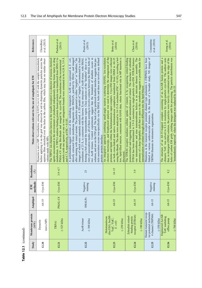
Table 12.1
(continued)
Study
Membrane protein
(MW)
Amphipol
EM
methods
Resolution
(Å)
Main observations relevant to the use of amphipols for EM
References
12.20
Dynactin
(not a MP)
A8-35
Cryo-EM
Dynactin is not a MP. Nevertheless, mixing 0.025% (w/v) A8-35 with the sample immediately
before applying it on the grid was observed to aid in dispersing dynactin particles across the
aqueous films stretched over the holes in the carbon film, which may bear on similar observations made with MPs.
Chowdhury
et al. (2015)
12.21
TRPA1
(~525 kDa)
PMAL-C8
Cryo-EM
3.9–4.7
The TRPA1 ion channel (also known as the wasabi receptor) is a detector of noxious chemical
agents encountered in the environment or produced endogenously during tissue injury or drug
metabolism. A TRPA1-maltose binding protein fusion protein was transferred from MNG-3 to
PMAL-C8 using Bio-Beads and imaged by cryo-EM. The resolutions of the reconstructed
models (with one agonist or one or two antagonists bound) were estimated to be 4.24 Å, 3.9 Å
and 4.7 Å, respectively, at FSC = 0.143.
Paulsen et al.
(2015)
12.22
AcrB trimer
(~360 kDa)
SMALPs
Negative
staining
23
A polyhistidine-tagged version of the multidrug transporter AcrB was expressed in E. coli,
directly extracted from membrane fragments into SMALPs, purified by IMAC, and imaged
after staining with uranyl acetate. The particles appeared as either "singlets" or "doublets," the
images of which were separately analyzed. A 3D model of "singlets." reconstructed to a final
resolution of 23 Å, is consistent with the known X-ray structure of AcrB trimer, with SMAL
forming a belt around the transmembrane region. Class analyses of the "doublets" show a variety of association modes, all of them seeming to involve contacts between the SMAL belts of
two AcrB trimers. Control experiments suggest that the formation of doublets occurs in
solution, and not on the EM grid. The lack of any preferred doublet interface suggests that particle association occurs non-specifically via the SMAL belts and does not involve any defined
protein–protein interface.
Postis et al.
(2015)
12.23
Holotranslocon
(SecYEG, SecDFYajC, YidC;
E. coli)
(~250 kDa)
A8-35
Cryo-EM
An integrative approach combining small-angle neutron scattering (SANS), low-resolution
electron microscopy, and biophysical analyses was used to determine the arrangement of the
proteins and lipids within the holotranslocon super-complex. Cryo-EM images were collected
on both cross-linked and native holotranslocon complexes transferred from DDM to A8-35
using Bio-Beads. The data were analyzed to 14-15-Å resolution (FSC = 0.143), revealing a hollow, lipid-filled interior, consistent with SANS data, whose functional role in MP insertion is
discussed..
Botte et al.
(2016)
12.24
Zebrafish retinolbinding protein
receptor (STRA6)
(~180 kDa)
A8-35
Cryo-EM
3.9
The STRA6 receptor mediates cellular uptake of vitamin A by recognizing retinol-binding
protein/retinol and triggering release and internalization of retinol. The structure of zebrafish
STRA6 has been determined to 3.9-Å resolution by single-particle cryo-EM. STRA6 features
one intramembrane and nine transmembrane helices in an intricate dimeric assembly. The
presence of a deep lipophilic cleft that is open to the membrane suggests a possible mode for
internalization of retinol through direct diffusion into the lipid bilayer.
Chen et al.
(2016)
12.25
Human transient receptor potential melastatin
4 channel (TRPM4)
(~550 kDa)
A8-35
Negative
staining
Mutations in the transient receptor potential channel type melastatin 4 (TRPM4) have been
linked to various cardiovascular diseases. In the frame of a broader study, NS images of
TRPM4 illustrate its tetrameric organization.
Constantine
et al. (2016)
12.26
Engineered AcrABTolC multidrug
efflux pump
(~760 kDa)
A8-35
Cryo-EM
8.2
The structure of an A8-35-trapped complex consisting of an AcrAB fusion protein and a
chimeric TolC was solved to 8.2-Å resolution by single-particle cryo-EM. The authors note
that, in detergent (DDM) solution, the particles were heavily concentrated near the edge of the
holes of carbon EM grids, yielding images unfit for processing. The particle distribution was
“tremendously improved” when the detergent was replaced by A8-35.
Jeong et al.
(2016)
14–15
12.3 The Use of Amphipols for Membrane Protein Electron Microscopy Studies
547
(continued)
Study
Membrane protein
(MW)
Amphipol
EM
methods
Resolution
(Å)
Main observations relevant to the use of amphipols for EM
References
12.20
Dynactin
(not a MP)
A8-35
Cryo-EM
Dynactin is not a MP. Nevertheless, mixing 0.025% (w/v) A8-35 with the sample immediately
before applying it on the grid was observed to aid in dispersing dynactin particles across the
aqueous films stretched over the holes in the carbon film, which may bear on similar observations made with MPs.
Chowdhury
et al. (2015)
12.21
TRPA1
(~525 kDa)
PMAL-C8
Cryo-EM
3.9–4.7
The TRPA1 ion channel (also known as the wasabi receptor) is a detector of noxious chemical
agents encountered in the environment or produced endogenously during tissue injury or drug
metabolism. A TRPA1-maltose binding protein fusion protein was transferred from MNG-3 to
PMAL-C8 using Bio-Beads and imaged by cryo-EM. The resolutions of the reconstructed
models (with one agonist or one or two antagonists bound) were estimated to be 4.24 Å, 3.9 Å
and 4.7 Å, respectively, at FSC = 0.143.
Paulsen et al.
(2015)
12.22
AcrB trimer
(~360 kDa)
SMALPs
Negative
staining
23
A polyhistidine-tagged version of the multidrug transporter AcrB was expressed in E. coli,
directly extracted from membrane fragments into SMALPs, purified by IMAC, and imaged
after staining with uranyl acetate. The particles appeared as either "singlets" or "doublets," the
images of which were separately analyzed. A 3D model of "singlets." reconstructed to a final
resolution of 23 Å, is consistent with the known X-ray structure of AcrB trimer, with SMAL
forming a belt around the transmembrane region. Class analyses of the "doublets" show a variety of association modes, all of them seeming to involve contacts between the SMAL belts of
two AcrB trimers. Control experiments suggest that the formation of doublets occurs in
solution, and not on the EM grid. The lack of any preferred doublet interface suggests that particle association occurs non-specifically via the SMAL belts and does not involve any defined
protein–protein interface.
Postis et al.
(2015)
12.23
Holotranslocon
(SecYEG, SecDFYajC, YidC;
E. coli)
(~250 kDa)
A8-35
Cryo-EM
An integrative approach combining small-angle neutron scattering (SANS), low-resolution
electron microscopy, and biophysical analyses was used to determine the arrangement of the
proteins and lipids within the holotranslocon super-complex. Cryo-EM images were collected
on both cross-linked and native holotranslocon complexes transferred from DDM to A8-35
using Bio-Beads. The data were analyzed to 14-15-Å resolution (FSC = 0.143), revealing a hollow, lipid-filled interior, consistent with SANS data, whose functional role in MP insertion is
discussed..
Botte et al.
(2016)
12.24
Zebrafish retinolbinding protein
receptor (STRA6)
(~180 kDa)
A8-35
Cryo-EM
3.9
The STRA6 receptor mediates cellular uptake of vitamin A by recognizing retinol-binding
protein/retinol and triggering release and internalization of retinol. The structure of zebrafish
STRA6 has been determined to 3.9-Å resolution by single-particle cryo-EM. STRA6 features
one intramembrane and nine transmembrane helices in an intricate dimeric assembly. The
presence of a deep lipophilic cleft that is open to the membrane suggests a possible mode for
internalization of retinol through direct diffusion into the lipid bilayer.
Chen et al.
(2016)
12.25
Human transient receptor potential melastatin
4 channel (TRPM4)
(~550 kDa)
A8-35
Negative
staining
Mutations in the transient receptor potential channel type melastatin 4 (TRPM4) have been
linked to various cardiovascular diseases. In the frame of a broader study, NS images of
TRPM4 illustrate its tetrameric organization.
Constantine
et al. (2016)
12.26
Engineered AcrABTolC multidrug
efflux pump
(~760 kDa)
A8-35
Cryo-EM
8.2
The structure of an A8-35-trapped complex consisting of an AcrAB fusion protein and a
chimeric TolC was solved to 8.2-Å resolution by single-particle cryo-EM. The authors note
that, in detergent (DDM) solution, the particles were heavily concentrated near the edge of the
holes of carbon EM grids, yielding images unfit for processing. The particle distribution was
“tremendously improved” when the detergent was replaced by A8-35.
Jeong et al.
(2016)
14–15
12.3 The Use of Amphipols for Membrane Protein Electron Microscopy Studies
547