

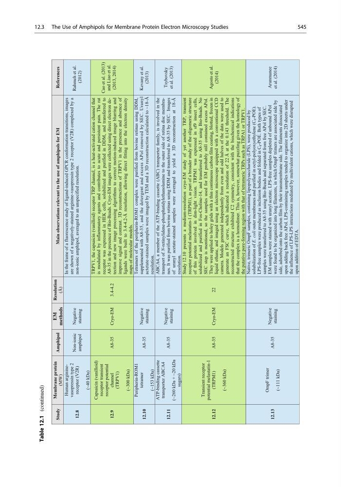
Table 12.1
(continued)
12.12
Transient receptor
potential melastatin-1
(TRPM1)
(~360 kDa)
A8-35
Cryo-EM
22
Study 12.10 presents a medium-resolution cryo-EM study of yet another TRP, transient
receptor potential melastatin-1 (TRPM1), as part of a broader study of the oligomeric structure
of this protein involved in dim-light vision. TRPM1 was expressed in sf9 insect cells,
solubilized and purified in Fos-Choline-14, and transferred to A8-35 using Bio-Beads. No
SEC step is mentioned, so the samples used for EM probably still contained excess APol.
They were applied to holey grids with a thin continuous carbon film coating, blotted, frozen in
liquid ethane, and imaged using an electron microscope equipped with a conventional CCD
camera. Models generated independently from even and odd halves of the data were used to
generate an FSC curve, which indicated a resolution of 22 Å at the 0.143 threshold. The
reconstructed structure exhibited C2 symmetry, consistent with the biochemical indications
that the complex is a homodimer. This is, however, somewhat puzzling given the homology of
the putative pore-forming region with that of tetrameric TRPs such as TRPA1 or TRPV1.
Agosto et al.
(2014)
12.13
OmpF trimer
(~111 kDa)
A8-35
Negative
staining
Native, trimeric OmpF samples, containing lipopolysaccharide (LPS), were produced by
solubilization of E. coli outer membranes and purified in octyl-polyoxyethylene (C
8 -POE).
LPS-free samples were produced as inclusion bodies and folded in C
8 -POE. Both types of
samples were transferred to A8-35 using Bio-Beads and separated from free APol by SEC.
EM samples were stained with uranyl acetate. LPS-free samples depleted of unbound APol
were found to be organized into long filaments, in which OmpF trimers are associated side by
side, adsorbed onto the carbon film by their transmembrane side. The filaments dissociated
upon adding back free APol. LPS-containing samples tended to organize into 2D sheets under
the influence of LPS-LPS interactions mediated by multivalent cations, which were disrupted
upon addition of EDTA.
Arunmanee
et al. (2014)
Study
Membrane protein
(MW)
Amphipol
EM
methods
Resolution
(Å)
Main observations relevant to the use of amphipols for EM
References
12.8
Human argininevasopressin type 2
receptor (V2R)
(~40 kDa)
Non-ionic
amphipol
Negative
staining
In the frame of a fluorescence study of ligand-induced GPCR conformation transitions, images
are shown of a negatively-stained arginine-vasopressin type 2 receptor (V2R) complexed by a
non-ionic amphipol, averaged to an unspecified resolution.
Rahmeh et al.
(2012)
12.9
Capsaicin (vanilloid)
receptor transient
receptor potential
channel
(TRPV1)
(~300 kDa)
A8-35
Cryo-EM
3.4-4.2
TRPV1, the capsaicin (vanilloid) receptor TRP channel, is a heat-activated cation channel that
is modulated by inflammatory agents and contributes to acute and persistent pain. The rat
receptor was expressed in HEK cells, solubilized and purified in DDM, and transferred to
A8-35 in the presence of Bio-Beads. Cryo-EM images were collected using direct electron detection and new image processing algorithms to correct motion-induced image blurring and
improve signal and contrast. 3D reconstructions of TRPV1 in the presence and absence of
ligands were built to 3.4-4.2-Å resolution, allowing direct fitting into the electron density
maps of molecular models.
Cao et al. (2013)
and Liao et al.
(2013, 2014)
12.10
Peripherin-ROM1
tetramer
(~153 kDa)
A8-35
Negative
staining
Tetramers of the peripherin-ROM1 complex were purified from bovine retinas using DDM,
supplemented with A8-35, and the detergent and excess APol removed by SEC. Uranyl
actetate-stained samples were imaged by TEM and a 3D reconstruction calculated to ~18-Å
resolution.
Kevany et al.
(2013)
12.11
ATP-binding cassette
transporter ABCA4
(~260 kDa + ~20 kDa
sugars)
A8-35
Negative
staining
ABCA4, a member of the ATP-binding cassette (ABC) transporter family, is involved in the
transport of N-retinylidene-phosphatidylethanolamine to the outer side of retina disc membranes. It was purified from bovine retinas using DDM and transferred to A8-35 by SEC. Images
of uranyl acetate-stained samples were averaged to yield a 3D reconstruction at 18-Å
resolution.
Tsybovsky
et al. (2013)
12.3 The Use of Amphipols for Membrane Protein Electron Microscopy Studies
545
(continued)
12.12
Transient receptor
potential melastatin-1
(TRPM1)
(~360 kDa)
A8-35
Cryo-EM
22
Study 12.10 presents a medium-resolution cryo-EM study of yet another TRP, transient
receptor potential melastatin-1 (TRPM1), as part of a broader study of the oligomeric structure
of this protein involved in dim-light vision. TRPM1 was expressed in sf9 insect cells,
solubilized and purified in Fos-Choline-14, and transferred to A8-35 using Bio-Beads. No
SEC step is mentioned, so the samples used for EM probably still contained excess APol.
They were applied to holey grids with a thin continuous carbon film coating, blotted, frozen in
liquid ethane, and imaged using an electron microscope equipped with a conventional CCD
camera. Models generated independently from even and odd halves of the data were used to
generate an FSC curve, which indicated a resolution of 22 Å at the 0.143 threshold. The
reconstructed structure exhibited C2 symmetry, consistent with the biochemical indications
that the complex is a homodimer. This is, however, somewhat puzzling given the homology of
the putative pore-forming region with that of tetrameric TRPs such as TRPA1 or TRPV1.
Agosto et al.
(2014)
12.13
OmpF trimer
(~111 kDa)
A8-35
Negative
staining
Native, trimeric OmpF samples, containing lipopolysaccharide (LPS), were produced by
solubilization of E. coli outer membranes and purified in octyl-polyoxyethylene (C
8 -POE).
LPS-free samples were produced as inclusion bodies and folded in C
8 -POE. Both types of
samples were transferred to A8-35 using Bio-Beads and separated from free APol by SEC.
EM samples were stained with uranyl acetate. LPS-free samples depleted of unbound APol
were found to be organized into long filaments, in which OmpF trimers are associated side by
side, adsorbed onto the carbon film by their transmembrane side. The filaments dissociated
upon adding back free APol. LPS-containing samples tended to organize into 2D sheets under
the influence of LPS-LPS interactions mediated by multivalent cations, which were disrupted
upon addition of EDTA.
Arunmanee
et al. (2014)
Study
Membrane protein
(MW)
Amphipol
EM
methods
Resolution
(Å)
Main observations relevant to the use of amphipols for EM
References
12.8
Human argininevasopressin type 2
receptor (V2R)
(~40 kDa)
Non-ionic
amphipol
Negative
staining
In the frame of a fluorescence study of ligand-induced GPCR conformation transitions, images
are shown of a negatively-stained arginine-vasopressin type 2 receptor (V2R) complexed by a
non-ionic amphipol, averaged to an unspecified resolution.
Rahmeh et al.
(2012)
12.9
Capsaicin (vanilloid)
receptor transient
receptor potential
channel
(TRPV1)
(~300 kDa)
A8-35
Cryo-EM
3.4-4.2
TRPV1, the capsaicin (vanilloid) receptor TRP channel, is a heat-activated cation channel that
is modulated by inflammatory agents and contributes to acute and persistent pain. The rat
receptor was expressed in HEK cells, solubilized and purified in DDM, and transferred to
A8-35 in the presence of Bio-Beads. Cryo-EM images were collected using direct electron detection and new image processing algorithms to correct motion-induced image blurring and
improve signal and contrast. 3D reconstructions of TRPV1 in the presence and absence of
ligands were built to 3.4-4.2-Å resolution, allowing direct fitting into the electron density
maps of molecular models.
Cao et al. (2013)
and Liao et al.
(2013, 2014)
12.10
Peripherin-ROM1
tetramer
(~153 kDa)
A8-35
Negative
staining
Tetramers of the peripherin-ROM1 complex were purified from bovine retinas using DDM,
supplemented with A8-35, and the detergent and excess APol removed by SEC. Uranyl
actetate-stained samples were imaged by TEM and a 3D reconstruction calculated to ~18-Å
resolution.
Kevany et al.
(2013)
12.11
ATP-binding cassette
transporter ABCA4
(~260 kDa + ~20 kDa
sugars)
A8-35
Negative
staining
ABCA4, a member of the ATP-binding cassette (ABC) transporter family, is involved in the
transport of N-retinylidene-phosphatidylethanolamine to the outer side of retina disc membranes. It was purified from bovine retinas using DDM and transferred to A8-35 by SEC. Images
of uranyl acetate-stained samples were averaged to yield a 3D reconstruction at 18-Å
resolution.
Tsybovsky
et al. (2013)
12.3 The Use of Amphipols for Membrane Protein Electron Microscopy Studies
545