

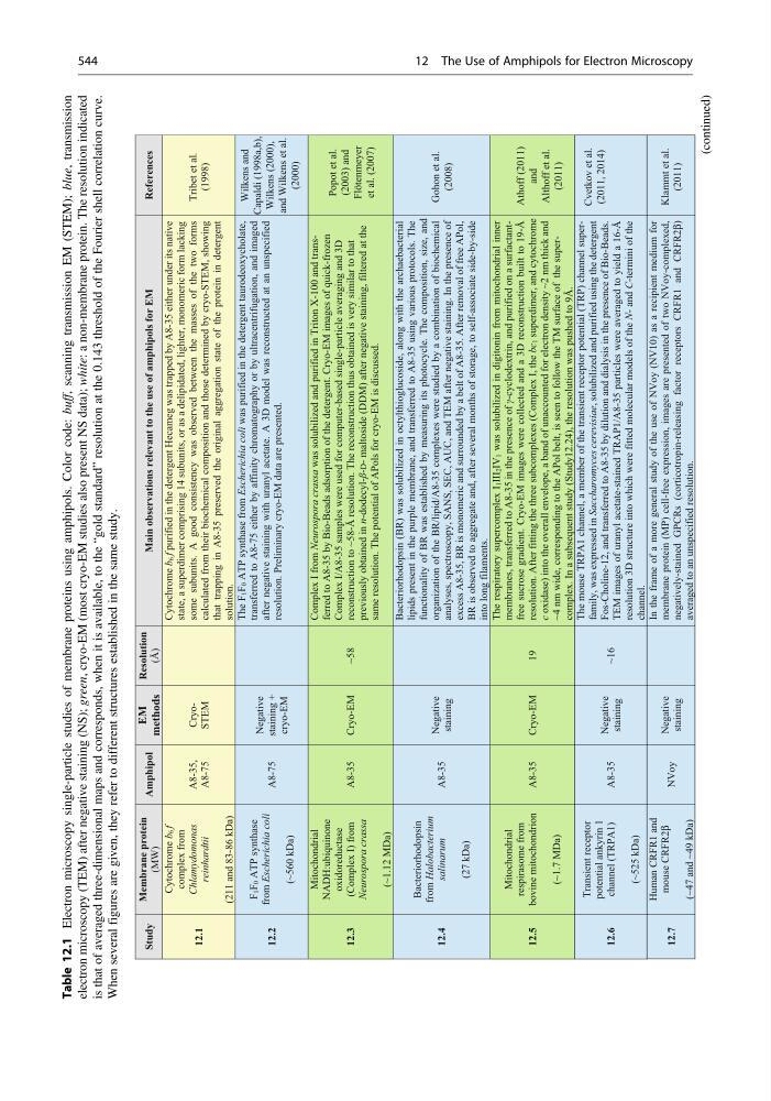
Table 12.1
Electron microscopy single-particle studies of membrane proteins using amphipols. Color code: buff,
scanning transmission EM (STEM); blue,
transmission
electron microscopy (TEM) after negative staining (NS); green,
cryo-EM (most cryo-EM studies also present NS data); white:
a non-membrane protein. The resolution indicated
is that of averaged three-dimensional maps and corresponds, when it is available, to the
“gold standard” resolution at the 0.143 threshold of the Fourier shell correlation curve.
When several figures are given, they refer to different structures established in the same study.
Study
Membrane protein
(MW)
Amphipol
EM
methods
Resolution
(Å)
Main observations relevant to the use of amphipols for EM
References
12.1
Cytochrome b6 f
complex from
Chlamydomonas
reinhardtii
(211 and 83-86 kDa)
A8-35,
A8-75
CryoSTEM
Cytochrome b6
f purified in the detergent Hecameg was trapped by A8-35 either under its native
state, a superdimer comprising 14 subunits, or as a delipidated, lighter, monomeric form lacking
some subunits. A good consistency was observed between the masses of the two forms
calculated from their biochemical composition and those determined by cryo-STEM, showing
that trapping in A8-35 preserved the original aggregation state of the protein in detergent
solution.
Tribet et al.
(1998)
12.2
F1F0
ATP synthase
from Escherichia coli
(~560 kDa)
A8-75
Negative
staining +
cryo-EM
The F1F0 ATP synthase from Escherichia coli was purified in the detergent taurodeoxycholate,
transferred to A8-75 either by affinity chromatography or by ultracentrifugation, and imaged
after negative staining with uranyl acetate. A 3D model was reconstructed at an unspecified
resolution. Preliminary cryo-EM data are presented.
Wilkens and
Capaldi (1998a,b),
Wilkens (2000),
and Wilkens et al.
(2000)
12.3
Mitochondrial
NADH:ubiquinone
oxidoreductase
(Complex I) from
Neurospora crassa
(~1.12 MDa)
A8-35
Cryo-EM
~58
Complex I from Neurospora crassa
was solubilized and purified in Triton X-100 and transferred to A8-35 by Bio-Beads adsorption of the detergent. Cryo-EM images of quick-frozen
Complex I/A8-35 samples were used for computer-based single-particle averaging and 3D
reconstruction to ~58-Å resolution. The reconstruction thus obtained is very similar to that
previously obtained in n-dodecyl-β-Dmaltoside (DDM) after negative staining, filtered at the
same resolution. The potential of APols for cryo-EM is discussed.
Popot et al.
(2003) and
Flötenmeyer
et al. (2007)
12.4
Bacteriorhodopsin
from
Halobacterium
salinarum
(27 kDa)
A8-35
Negative
staining
Bacteriorhodopsin (BR) was solubilized in octylthioglucoside, along with the archaebacterial
lipids present in the purple membrane, and transferred to A8-35 using various protocols. The
functionality of BR was established by measuring its photocycle. The composition, size, and
organization of the BR/lipid/A8-35 complexes were studied by a combination of biochemical
analyses, spectroscopy, SANS, SEC, AUC, and TEM after negative staining. In the presence of
excess A8-35, BR is monomeric and surrounded by a belt of A8-35. After removal of free APol,
BR is observed to aggregate and, after several months of storage,
to self-associate side-by-side
into long filaments.
Gohon et al.
(2008)
12.5
Mitochondrial
respirasome from
bovine mitochondrion
(~1.7 MDa)
A8-35
Cryo-EM
19
The respiratory supercomplex I1III2IV1 was solubilized in digitonin from mitochondrial inner
membranes, transferred to A8-35 in the presence of γ-cyclodextrin, and purified on a surfactantfree sucrose gradient. Cryo-EM images were collected and a 3D reconstruction
built to 19-Å
resolution. After fitting the three subcomplexes (Complex I, the bc1 superdimer, and cytochrome
c oxidase) into the overall envelope, a band of unaccounted for electron density ~2 nm thick and
~4 nm wide, corresponding to the APol belt, is seen to follow the TM surface
of the supercomplex. In a subsequent study (Study12.24), the resolution was pushed to 9Å.
Athoff (2011)
and
Althoff et al.
(2011)
12.6
Transient receptor
potential ankyrin 1
channel (TRPA1)
(~525 kDa)
A8-35
Negative
staining
~16
The mouse TRPA1 channel, a member of the transient receptor potential (TRP) channel superfamily, was expressed in Saccharomyces cerevisiae, solubilized and purified using the detergent
Fos-Choline-12, and transferred to A8-35 by dilution and dialysis in the presence of Bio-Beads.
TEM images of uranyl acetate-stained TRAP1/A8-35 particles were averaged to yield a 16-Å
resolution 3D structure into which were fitted molecular models of the N- and C-termini of the
channel.
Cvetkov et al.
(2011, 2014)
12.7
Human CRFR1 and
mouse CRFR2β
(~47 and ~49 kDa)
NVoy
Negative
staining
In the frame of a more general study of the use of NVoy (NV10) as a recipient medium for
membrane protein (MP) cell-free expression, images are presented of two NVoy-complexed,
negatively-stained GPCRs (corticotropin-releasing factor receptors CRFR1 and CRFR2β)
averaged to an unspecified resolution.
Klammt et al.
(2011)
(continued)
544
12 The Use of Amphipols for Electron Microscopy
Electron microscopy single-particle studies of membrane proteins using amphipols. Color code: buff,
scanning transmission EM (STEM); blue,
transmission
electron microscopy (TEM) after negative staining (NS); green,
cryo-EM (most cryo-EM studies also present NS data); white:
a non-membrane protein. The resolution indicated
is that of averaged three-dimensional maps and corresponds, when it is available, to the
“gold standard” resolution at the 0.143 threshold of the Fourier shell correlation curve.
When several figures are given, they refer to different structures established in the same study.
Study
Membrane protein
(MW)
Amphipol
EM
methods
Resolution
(Å)
Main observations relevant to the use of amphipols for EM
References
12.1
Cytochrome b6 f
complex from
Chlamydomonas
reinhardtii
(211 and 83-86 kDa)
A8-35,
A8-75
CryoSTEM
Cytochrome b6
f purified in the detergent Hecameg was trapped by A8-35 either under its native
state, a superdimer comprising 14 subunits, or as a delipidated, lighter, monomeric form lacking
some subunits. A good consistency was observed between the masses of the two forms
calculated from their biochemical composition and those determined by cryo-STEM, showing
that trapping in A8-35 preserved the original aggregation state of the protein in detergent
solution.
Tribet et al.
(1998)
12.2
F1F0
ATP synthase
from Escherichia coli
(~560 kDa)
A8-75
Negative
staining +
cryo-EM
The F1F0 ATP synthase from Escherichia coli was purified in the detergent taurodeoxycholate,
transferred to A8-75 either by affinity chromatography or by ultracentrifugation, and imaged
after negative staining with uranyl acetate. A 3D model was reconstructed at an unspecified
resolution. Preliminary cryo-EM data are presented.
Wilkens and
Capaldi (1998a,b),
Wilkens (2000),
and Wilkens et al.
(2000)
12.3
Mitochondrial
NADH:ubiquinone
oxidoreductase
(Complex I) from
Neurospora crassa
(~1.12 MDa)
A8-35
Cryo-EM
~58
Complex I from Neurospora crassa
was solubilized and purified in Triton X-100 and transferred to A8-35 by Bio-Beads adsorption of the detergent. Cryo-EM images of quick-frozen
Complex I/A8-35 samples were used for computer-based single-particle averaging and 3D
reconstruction to ~58-Å resolution. The reconstruction thus obtained is very similar to that
previously obtained in n-dodecyl-β-Dmaltoside (DDM) after negative staining, filtered at the
same resolution. The potential of APols for cryo-EM is discussed.
Popot et al.
(2003) and
Flötenmeyer
et al. (2007)
12.4
Bacteriorhodopsin
from
Halobacterium
salinarum
(27 kDa)
A8-35
Negative
staining
Bacteriorhodopsin (BR) was solubilized in octylthioglucoside, along with the archaebacterial
lipids present in the purple membrane, and transferred to A8-35 using various protocols. The
functionality of BR was established by measuring its photocycle. The composition, size, and
organization of the BR/lipid/A8-35 complexes were studied by a combination of biochemical
analyses, spectroscopy, SANS, SEC, AUC, and TEM after negative staining. In the presence of
excess A8-35, BR is monomeric and surrounded by a belt of A8-35. After removal of free APol,
BR is observed to aggregate and, after several months of storage,
to self-associate side-by-side
into long filaments.
Gohon et al.
(2008)
12.5
Mitochondrial
respirasome from
bovine mitochondrion
(~1.7 MDa)
A8-35
Cryo-EM
19
The respiratory supercomplex I1III2IV1 was solubilized in digitonin from mitochondrial inner
membranes, transferred to A8-35 in the presence of γ-cyclodextrin, and purified on a surfactantfree sucrose gradient. Cryo-EM images were collected and a 3D reconstruction
built to 19-Å
resolution. After fitting the three subcomplexes (Complex I, the bc1 superdimer, and cytochrome
c oxidase) into the overall envelope, a band of unaccounted for electron density ~2 nm thick and
~4 nm wide, corresponding to the APol belt, is seen to follow the TM surface
of the supercomplex. In a subsequent study (Study12.24), the resolution was pushed to 9Å.
Athoff (2011)
and
Althoff et al.
(2011)
12.6
Transient receptor
potential ankyrin 1
channel (TRPA1)
(~525 kDa)
A8-35
Negative
staining
~16
The mouse TRPA1 channel, a member of the transient receptor potential (TRP) channel superfamily, was expressed in Saccharomyces cerevisiae, solubilized and purified using the detergent
Fos-Choline-12, and transferred to A8-35 by dilution and dialysis in the presence of Bio-Beads.
TEM images of uranyl acetate-stained TRAP1/A8-35 particles were averaged to yield a 16-Å
resolution 3D structure into which were fitted molecular models of the N- and C-termini of the
channel.
Cvetkov et al.
(2011, 2014)
12.7
Human CRFR1 and
mouse CRFR2β
(~47 and ~49 kDa)
NVoy
Negative
staining
In the frame of a more general study of the use of NVoy (NV10) as a recipient medium for
membrane protein (MP) cell-free expression, images are presented of two NVoy-complexed,
negatively-stained GPCRs (corticotropin-releasing factor receptors CRFR1 and CRFR2β)
averaged to an unspecified resolution.
Klammt et al.
(2011)
(continued)
544
12 The Use of Amphipols for Electron Microscopy